The structure of hyperthermophilic beta-N-acetylglucosaminidase reveals a novel dimer architecture associated with the active site.
Mine, S., Kado, Y., Watanabe, M., Fukuda, Y., Abe, Y., Ueda, T., Kawarabayasi, Y., Inoue, T., Ishikawa, K.(2014) FEBS J 281: 5092-5103
- PubMed: 25227262
- DOI: https://doi.org/10.1111/febs.13049
- Primary Citation of Related Structures:
3WO8 - PubMed Abstract:
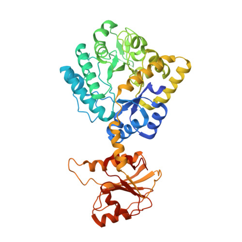
The β-N-acetylglucosaminidase from the hyperthermophilic bacteria Thermotoga maritima (NagA) hydrolyzes chitooligomers into monomer β-N-acetylglucosamine. Although NagA contains a highly conserved sequence motif found in glycoside hydrolase (GH) family 3, it can be distinguished from other GH family 3 β-N-acetylglucosaminidases by its substrate specificity and biological assembly. To investigate its unique structure around the active site, we determined the crystal structure of NagA at a resolution of 2.43 Å. The NagA forms a dimer structure in which the monomer structure consists of an N- and a C-terminal domain. The dimer structure exhibits high solvation free energy for dimer formation. From mutagenesis analyses, the catalytic nucleophile and general acid-base residues were supposed to be Asp245 and His173, respectively. The most striking characteristic of NagA was that it forms the active site cleft from the N-terminal domain and the C-terminal domain of the next polypeptide chain, whereas the other two-domain GH family 3 enzymes form the site within the same molecule. Another striking feature is that the loops located around the active site show high flexibility. One of the flexible loops contains the general acid-base His173 and was thought to be involved in substrate distortion during catalysis. In addition, a loop in close contact with the active site, which comes from the C-terminal domain of the next polypeptide chain, contains a region of high B-factor values, indicating the possibility that the C-terminal domain is involved in catalysis. These results suggest that the dimer structure of NagA is important for its activity and thermostability.
Organizational Affiliation:
National Institute of Advanced Industrial Science and Technology (AIST), Hyogo, Japan.