3WIQ

Crystal structure of kojibiose phosphorylase complexed with kojibiose


Experimental Data Snapshot

  • Method: X-RAY DIFFRACTION
  • Resolution: 2.80 Å
  • R-Value Free: 0.259 
  • R-Value Work: 0.204 
  • R-Value Observed: 0.207 

wwPDB Validation   3D Report Full Report


This is version 2.1 of the entry. See complete history


Literature

Structural and mutational analysis of substrate recognition in kojibiose phosphorylase

Okada, S.Yamamoto, T.Watanabe, H.Nishimoto, T.Chaen, H.Fukuda, S.Wakagi, T.Fushinobu, S.

(2014) FEBS J 281: 778-786

  • DOI: https://doi.org/10.1111/febs.12622
  • Primary Citation of Related Structures:  
    3WIQ, 3WIR

  • PubMed Abstract: 

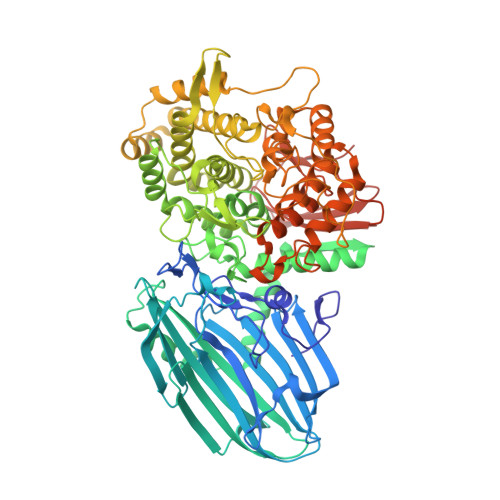
    Glycoside hydrolase (GH) family 65 contains phosphorylases acting on maltose (Glc-α1,4-Glc), kojibiose (Glc-α1,2-Glc), trehalose (Glc-α1,α1,-Glc), and nigerose (Glc-α1,3-Glc). These phosphorylases can efficiently catalyze the reverse reactions with high specificities, and thus can be applied to the practical synthesis of α-glucosyl oligosaccharides. Here, we determined the crystal structures of kojibiose phosphorylase from Caldicellulosiruptor saccharolyticus in complex with glucose and phosphate and in complex with kojibiose and sulfate, providing the first structural insights into the substrate recognition of a glycoside hydrolase family 65 enzyme. The loop 3 region comprising the active site of kojibiose phosphorylase is significantly longer than the active sites of other enzymes, and three residues around this loop, Trp391, Glu392, and Thr417, recognize kojibiose. Various mutants mimicking the residue conservation patterns of other phosphorylases were constructed by mutation at these three residues. Activity measurements of the mutants against four substrates indicated that Trp391 and Glu392, especially the latter, are required for the kojibiose activity.


  • Organizational Affiliation

    Department of Biotechnology, University of Tokyo, Japan.


Macromolecules
Find similar proteins by:  (by identity cutoff)  |  3D Structure
Entity ID: 1
MoleculeChains Sequence LengthOrganismDetailsImage
Kojibiose phosphorylase764Caldicellulosiruptor saccharolyticus DSM 8903Mutation(s): 0 
Gene Names: Csac_0444
EC: 2.4.1.230
UniProt
Find proteins for A4XGP2 (Caldicellulosiruptor saccharolyticus (strain ATCC 43494 / DSM 8903 / Tp8T 6331))
Explore A4XGP2 
Go to UniProtKB:  A4XGP2
Entity Groups  
Sequence Clusters30% Identity50% Identity70% Identity90% Identity95% Identity100% Identity
UniProt GroupA4XGP2
Sequence Annotations
Expand
  • Reference Sequence
Oligosaccharides

Help

Entity ID: 2
MoleculeChains Length2D Diagram Glycosylation3D Interactions
alpha-D-glucopyranose-(1-2)-beta-D-glucopyranose
B
2N/A
Glycosylation Resources
GlyTouCan:  G83663CO
GlyCosmos:  G83663CO
Small Molecules
Ligands 1 Unique
IDChains Name / Formula / InChI Key2D Diagram3D Interactions
SO4
Query on SO4

Download Ideal Coordinates CCD File 
C [auth A]SULFATE ION
O4 S
QAOWNCQODCNURD-UHFFFAOYSA-L
Experimental Data & Validation

Experimental Data

  • Method: X-RAY DIFFRACTION
  • Resolution: 2.80 Å
  • R-Value Free: 0.259 
  • R-Value Work: 0.204 
  • R-Value Observed: 0.207 
  • Space Group: I 4 2 2
Unit Cell:
Length ( Å )Angle ( ˚ )
a = 192.506α = 90
b = 192.506β = 90
c = 202.221γ = 90
Software Package:
Software NamePurpose
HKL-2000data collection
MOLREPphasing
REFMACrefinement
HKL-2000data reduction
HKL-2000data scaling

Structure Validation

View Full Validation Report


Entry History 

Deposition Data

Revision History  (Full details and data files)

  • Version 1.0: 2014-02-05
    Type: Initial release
  • Version 2.0: 2020-07-29
    Type: Remediation
    Reason: Carbohydrate remediation
    Changes: Atomic model, Data collection, Database references, Derived calculations, Structure summary
  • Version 2.1: 2023-11-08
    Changes: Data collection, Database references, Refinement description, Structure summary