3WCS

Crystal structure of plant lectin (ligand-bound form)


Experimental Data Snapshot

  • Method: X-RAY DIFFRACTION
  • Resolution: 1.75 Å
  • R-Value Free: 0.255 
  • R-Value Work: 0.237 
  • R-Value Observed: 0.238 

wwPDB Validation   3D Report Full Report


Ligand Structure Quality Assessment 


This is version 2.1 of the entry. See complete history


Literature

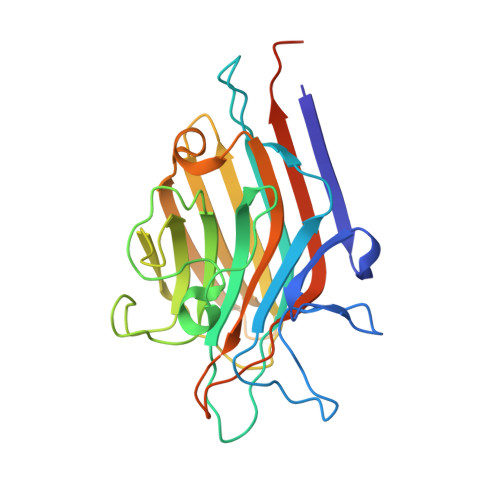
Phytohemagglutinin from Phaseolus vulgaris (PHA-E) displays a novel glycan recognition mode using a common legume lectin fold

Nagae, M.Soga, K.Morita-Matsumoto, K.Hanashima, S.Ikeda, A.Yamamoto, K.Yamaguchi, Y.

(2014) Glycobiology 24: 368-378

  • DOI: https://doi.org/10.1093/glycob/cwu004
  • Primary Citation of Related Structures:  
    3WCR, 3WCS, 3WOG

  • PubMed Abstract: 

    Phytohemagglutinin from Phaseolus vulgaris (PHA-E), a legume lectin, has an unusual specificity toward biantennary galactosylated N-glycan with bisecting N-acetylglucosamine (GlcNAc). To investigate the interaction in detail, we have solved the crystal structures of PHA-E without ligand and in complex with biantennary N-glycan derivatives. PHA-E interacts with the trisaccharide unit (Galβ1-4GlcNAcβ1-2Man) in a manner completely different from that of mannose/glucose-specific legume lectins. The inner mannose residue binds to a novel site on the protein, and its rotation is opposite to that occurring in the monosaccharide-binding site of other lectins around the sugar O3 axis. Saturation-transfer difference NMR using biantennary di-galactosylated and bisected glycans reveals that PHA-E interacts with both antennas almost equally. The unique carbohydrate interaction explains the glycan-binding specificity and high affinity.


  • Organizational Affiliation

    Structural Glycobiology Team, Systems Glycobiology Research Group, RIKEN-Max Planck Joint Research Center, RIKEN Global Research Cluster, 2-1 Hirosawa, Wako, Saitama 351-0198, Japan.


Macromolecules
Find similar proteins by:  (by identity cutoff)  |  3D Structure
Entity ID: 1
MoleculeChains Sequence LengthOrganismDetailsImage
Erythroagglutinin
A, B
254Phaseolus vulgarisMutation(s): 0 
UniProt
Find proteins for P05088 (Phaseolus vulgaris)
Explore P05088 
Go to UniProtKB:  P05088
Entity Groups  
Sequence Clusters30% Identity50% Identity70% Identity90% Identity95% Identity100% Identity
UniProt GroupP05088
Sequence Annotations
Expand
  • Reference Sequence
Oligosaccharides

Help

Entity ID: 2
MoleculeChains Length2D Diagram Glycosylation3D Interactions
beta-D-galactopyranose-(1-4)-2-acetamido-2-deoxy-beta-D-glucopyranose-(1-2)-alpha-D-mannopyranose
C
3N/A
Glycosylation Resources
GlyTouCan:  G28052FT
GlyCosmos:  G28052FT
GlyGen:  G28052FT
Entity ID: 3
MoleculeChains Length2D Diagram Glycosylation3D Interactions
2-acetamido-2-deoxy-beta-D-glucopyranose-(1-2)-alpha-D-mannopyranose
D
2N/A
Glycosylation Resources
GlyTouCan:  G85856KC
GlyCosmos:  G85856KC
GlyGen:  G85856KC
Small Molecules
Ligands 4 Unique
IDChains Name / Formula / InChI Key2D Diagram3D Interactions
NAG
Query on NAG

Download Ideal Coordinates CCD File 
E [auth A],
M [auth B]
2-acetamido-2-deoxy-beta-D-glucopyranose
C8 H15 N O6
OVRNDRQMDRJTHS-FMDGEEDCSA-N
EDO
Query on EDO

Download Ideal Coordinates CCD File 
H [auth A],
I [auth A],
J [auth A],
K [auth A],
L [auth A]
1,2-ETHANEDIOL
C2 H6 O2
LYCAIKOWRPUZTN-UHFFFAOYSA-N
MN
Query on MN

Download Ideal Coordinates CCD File 
F [auth A],
N [auth B]
MANGANESE (II) ION
Mn
WAEMQWOKJMHJLA-UHFFFAOYSA-N
CA
Query on CA

Download Ideal Coordinates CCD File 
G [auth A],
O [auth B]
CALCIUM ION
Ca
BHPQYMZQTOCNFJ-UHFFFAOYSA-N
Experimental Data & Validation

Experimental Data

  • Method: X-RAY DIFFRACTION
  • Resolution: 1.75 Å
  • R-Value Free: 0.255 
  • R-Value Work: 0.237 
  • R-Value Observed: 0.238 
  • Space Group: C 2 2 21
Unit Cell:
Length ( Å )Angle ( ˚ )
a = 76.579α = 90
b = 196.193β = 90
c = 98.655γ = 90
Software Package:
Software NamePurpose
SERGUIdata collection
MOLREPphasing
REFMACrefinement
HKL-2000data reduction
HKL-2000data scaling

Structure Validation

View Full Validation Report


Ligand Structure Quality Assessment 


Entry History 

Deposition Data

Revision History  (Full details and data files)

  • Version 1.0: 2014-04-09
    Type: Initial release
  • Version 1.1: 2017-11-22
    Changes: Refinement description
  • Version 2.0: 2020-07-29
    Type: Remediation
    Reason: Carbohydrate remediation
    Changes: Atomic model, Data collection, Derived calculations, Structure summary
  • Version 2.1: 2023-11-08
    Changes: Data collection, Database references, Refinement description, Structure summary