3WCK

Crystal structure of monomeric photosensitizing fluorescent protein, Supernova


Experimental Data Snapshot

  • Method: X-RAY DIFFRACTION
  • Resolution: 2.30 Å
  • R-Value Free: 0.234 
  • R-Value Work: 0.181 
  • R-Value Observed: 0.184 

wwPDB Validation   3D Report Full Report


This is version 2.0 of the entry. See complete history


Literature

SuperNova, a monomeric photosensitizing fluorescent protein for chromophore-assisted light inactivation

Takemoto, K.Matsuda, T.Sakai, N.Fu, D.Noda, M.Uchiyama, S.Kotera, I.Arai, Y.Horiuchi, M.Fukui, K.Ayabe, T.Inagaki, F.Suzuki, H.Nagai, T.

(2013) Sci Rep 3: 2629-2629

  • DOI: https://doi.org/10.1038/srep02629
  • Primary Citation of Related Structures:  
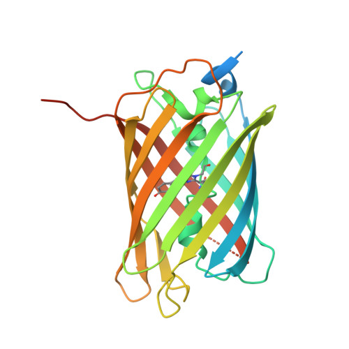
    3WCK

  • PubMed Abstract: 

    Chromophore-assisted light inactivation (CALI) is a powerful technique for acute perturbation of biomolecules in a spatio-temporally defined manner in living specimen with reactive oxygen species (ROS). Whereas a chemical photosensitizer including fluorescein must be added to specimens exogenously and cannot be restricted to particular cells or sub-cellular compartments, a genetically-encoded photosensitizer, KillerRed, can be controlled in its expression by tissue specific promoters or subcellular localization tags. Despite of this superiority, KillerRed hasn't yet become a versatile tool because its dimerization tendency prevents fusion with proteins of interest. Here, we report the development of monomeric variant of KillerRed (SuperNova) by direct evolution using random mutagenesis. In contrast to KillerRed, SuperNova in fusion with target proteins shows proper localization. Furthermore, unlike KillerRed, SuperNova expression alone doesn't perturb mitotic cell division. Supernova retains the ability to generate ROS, and hence promote CALI-based functional analysis of target proteins overcoming the major drawbacks of KillerRed.


  • Organizational Affiliation

    1] Research Institute for Electronic Sciences, Hokkaido University, Kita-20 Nishi-10 Kita-ku, Sapporo, Hokkaido 001-0020, Japan [2] [3].


Macromolecules
Find similar proteins by:  (by identity cutoff)  |  3D Structure
Entity ID: 1
MoleculeChains Sequence LengthOrganismDetailsImage
Monomeric photosenitizing fluorescent protein supernova
A, B, C, D, E
A, B, C, D, E, F, G, H
271synthetic constructMutation(s): 1 
UniProt
Find proteins for Q2TCH5 (Anthomedusae sp. DC-2005)
Explore Q2TCH5 
Go to UniProtKB:  Q2TCH5
Entity Groups  
Sequence Clusters30% Identity50% Identity70% Identity90% Identity95% Identity100% Identity
UniProt GroupQ2TCH5
Sequence Annotations
Expand
  • Reference Sequence
Small Molecules
Modified Residues  1 Unique
IDChains TypeFormula2D DiagramParent
CRQ
Query on CRQ
A, B, C, D, E
A, B, C, D, E, F, G, H
L-PEPTIDE LINKINGC16 H16 N4 O5GLN, TYR, GLY
Experimental Data & Validation

Experimental Data

  • Method: X-RAY DIFFRACTION
  • Resolution: 2.30 Å
  • R-Value Free: 0.234 
  • R-Value Work: 0.181 
  • R-Value Observed: 0.184 
  • Space Group: P 1
Unit Cell:
Length ( Å )Angle ( ˚ )
a = 60.252α = 70.57
b = 89.304β = 89.95
c = 109.885γ = 71.62
Software Package:
Software NamePurpose
PHASERphasing
REFMACrefinement
HKL-2000data reduction
HKL-2000data scaling

Structure Validation

View Full Validation Report


Entry History 

Deposition Data

Revision History  (Full details and data files)

  • Version 1.0: 2013-10-02
    Type: Initial release
  • Version 1.1: 2023-11-08
    Changes: Data collection, Database references, Derived calculations, Refinement description
  • Version 2.0: 2023-11-15
    Changes: Atomic model, Data collection, Derived calculations