3W9A

Crystal structure of the catalytic domain of the glycoside hydrolase family 131 protein from Coprinopsis cinerea


Experimental Data Snapshot

  • Method: X-RAY DIFFRACTION
  • Resolution: 1.99 Å
  • R-Value Free: 0.236 
  • R-Value Work: 0.188 
  • R-Value Observed: 0.191 

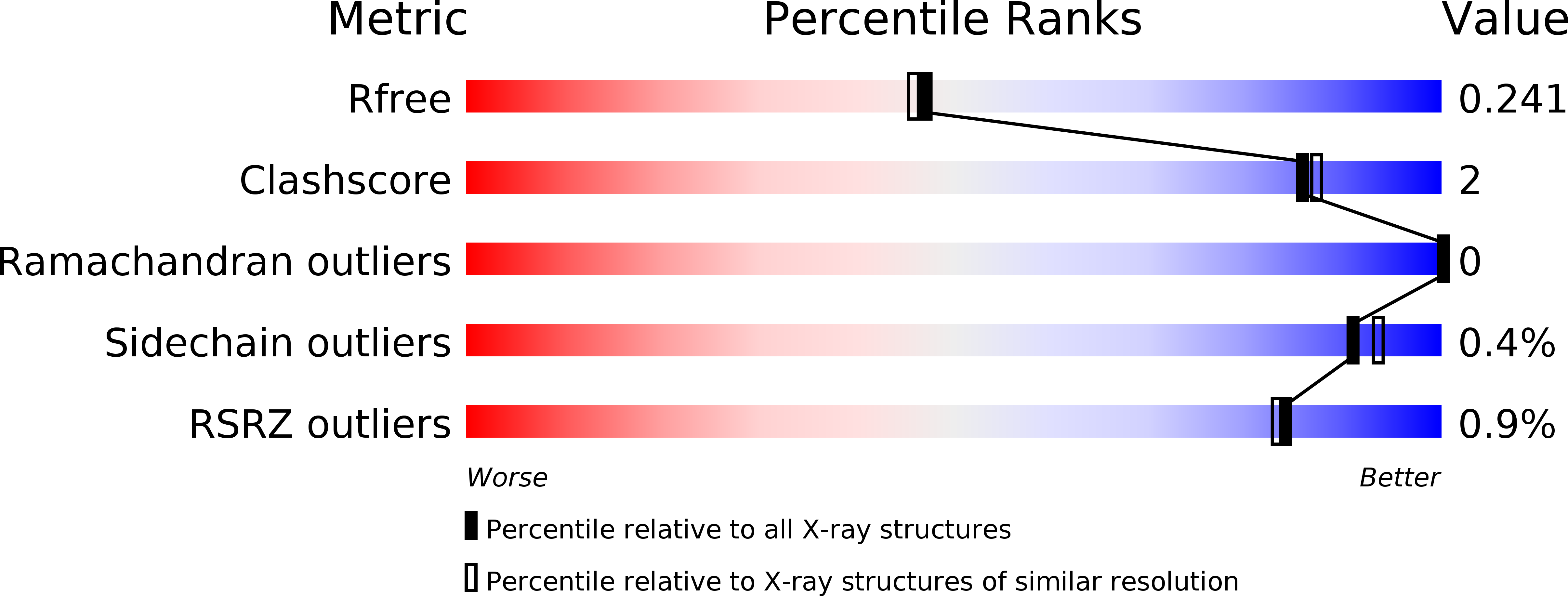
wwPDB Validation   3D Report Full Report


This is version 1.2 of the entry. See complete history


Literature

Crystal structure of the N-terminal domain of a glycoside hydrolase family 131 protein from Coprinopsis cinerea

Miyazaki, T.Yoshida, M.Tamura, M.Tanaka, Y.Umezawa, K.Nishikawa, A.Tonozuka, T.

(2013) FEBS Lett 587: 2193-2198

  • DOI: https://doi.org/10.1016/j.febslet.2013.05.041
  • Primary Citation of Related Structures:  
    3W9A

  • PubMed Abstract: 

    The crystal structure of the N-terminal putative catalytic domain of a glycoside hydrolase family 131 protein from Coprinopsis cinerea (CcGH131A) was determined. The structure of CcGH131A was found to be composed of a β-jelly roll fold and mainly consisted of two β-sheets, sheet-A and sheet-B. A concave of sheet-B, the possible active site, was wide and shallow, and three glycerol molecules were present in the concave. Arg96, Glu98, Glu138, and His218 are likely to be catalytically critical residues, and it was suggested that the catalytic mechanism of CcGH131A is different from that of typical glycosidases.


  • Organizational Affiliation

    Department of Applied Biological Science, Tokyo University of Agriculture and Technology, Tokyo 183-8509, Japan.


Macromolecules
Find similar proteins by:  (by identity cutoff)  |  3D Structure
Entity ID: 1
MoleculeChains Sequence LengthOrganismDetailsImage
Putative uncharacterized protein
A, B, C, D
245Coprinopsis cinerea okayama7#130Mutation(s): 0 
Gene Names: CC1G_07166
UniProt
Find proteins for A8NRB3 (Coprinopsis cinerea (strain Okayama-7 / 130 / ATCC MYA-4618 / FGSC 9003))
Explore A8NRB3 
Go to UniProtKB:  A8NRB3
Entity Groups  
Sequence Clusters30% Identity50% Identity70% Identity90% Identity95% Identity100% Identity
UniProt GroupA8NRB3
Sequence Annotations
Expand
  • Reference Sequence
Small Molecules
Ligands 1 Unique
IDChains Name / Formula / InChI Key2D Diagram3D Interactions
GOL
Query on GOL

Download Ideal Coordinates CCD File 
E [auth A]
F [auth A]
G [auth A]
H [auth B]
I [auth B]
E [auth A],
F [auth A],
G [auth A],
H [auth B],
I [auth B],
J [auth B],
K [auth C],
L [auth C],
M [auth C],
N [auth D],
O [auth D],
P [auth D]
GLYCEROL
C3 H8 O3
PEDCQBHIVMGVHV-UHFFFAOYSA-N
Modified Residues  1 Unique
IDChains TypeFormula2D DiagramParent
MSE
Query on MSE
A, B, C, D
L-PEPTIDE LINKINGC5 H11 N O2 SeMET
Experimental Data & Validation

Experimental Data

  • Method: X-RAY DIFFRACTION
  • Resolution: 1.99 Å
  • R-Value Free: 0.236 
  • R-Value Work: 0.188 
  • R-Value Observed: 0.191 
  • Space Group: P 1
Unit Cell:
Length ( Å )Angle ( ˚ )
a = 59.2α = 89.99
b = 68.421β = 72.87
c = 69.168γ = 85.81
Software Package:
Software NamePurpose
ADSCdata collection
AutoSolphasing
REFMACrefinement
HKL-2000data reduction
HKL-2000data scaling

Structure Validation

View Full Validation Report


Entry History 

Deposition Data

Revision History  (Full details and data files)

  • Version 1.0: 2013-05-22
    Type: Initial release
  • Version 1.1: 2013-06-05
    Changes: Database references
  • Version 1.2: 2013-07-10
    Changes: Database references