3VW5

Crystal structure of sugar epimerase from ruminal bacterium


Experimental Data Snapshot

  • Method: X-RAY DIFFRACTION
  • Resolution: 2.60 Å
  • R-Value Free: 0.222 
  • R-Value Work: 0.173 
  • R-Value Observed: 0.175 

wwPDB Validation   3D Report Full Report


This is version 1.1 of the entry. See complete history


Literature

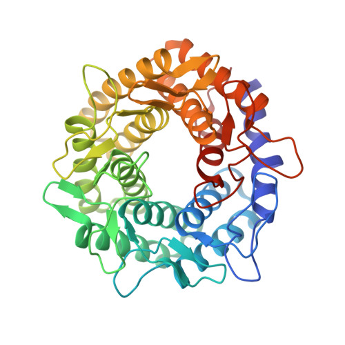
Crystal structure of Ruminococcus albus cellobiose 2-epimerase: structural insights into epimerization of unmodified sugar

Fujiwara, T.Saburi, W.Inoue, S.Mori, H.Matsui, H.Tanaka, I.Yao, M.

(2013) FEBS Lett 587: 840-846

  • DOI: https://doi.org/10.1016/j.febslet.2013.02.007
  • Primary Citation of Related Structures:  
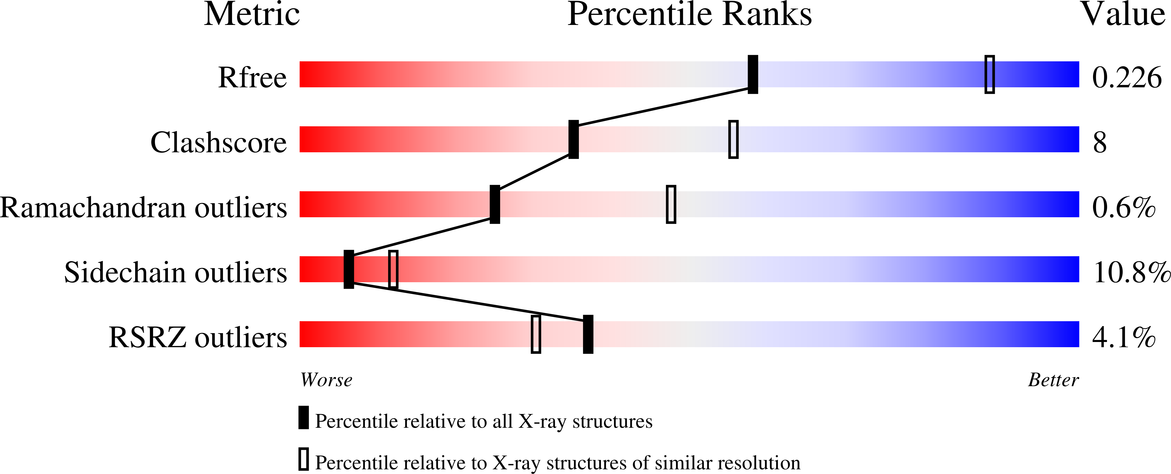
    3VW5

  • PubMed Abstract: 

    Enzymatic epimerization is an important modification for carbohydrates to acquire diverse functions attributable to their stereoisomers. Cellobiose 2-epimerase (CE) catalyzes interconversion between d-glucose and d-mannose residues at the reducing end of β-1,4-linked oligosaccharides. Here, we solved the structure of Ruminococcus albus CE (RaCE). The structure of RaCE showed strong similarity to those of N-acetyl-D-glucosamine 2-epimerase and aldose-ketose isomerase YihS with a high degree of conservation of residues around the catalytic center, although sequence identity between them is low. Based on structural comparison, we found that His184 is required for RaCE activity as the third histidine added to two essential histidines in other sugar epimerases/isomerases. This finding was confirmed by mutagenesis, suggesting a new catalytic mechanism for CE involving three histidines.


  • Organizational Affiliation

    Graduate School of Life Science, Hokkaido University, Kita-ku, Sapporo, Japan.


Macromolecules
Find similar proteins by:  (by identity cutoff)  |  3D Structure
Entity ID: 1
MoleculeChains Sequence LengthOrganismDetailsImage
Cellobiose 2-epimerase
A, B, C
389Ruminococcus albusMutation(s): 2 
Gene Names: CE
EC: 5.1.3.11
UniProt
Find proteins for P0DKY4 (Ruminococcus albus)
Explore P0DKY4 
Go to UniProtKB:  P0DKY4
Entity Groups  
Sequence Clusters30% Identity50% Identity70% Identity90% Identity95% Identity100% Identity
UniProt GroupP0DKY4
Sequence Annotations
Expand
  • Reference Sequence
Experimental Data & Validation

Experimental Data

  • Method: X-RAY DIFFRACTION
  • Resolution: 2.60 Å
  • R-Value Free: 0.222 
  • R-Value Work: 0.173 
  • R-Value Observed: 0.175 
  • Space Group: P 21 21 21
Unit Cell:
Length ( Å )Angle ( ˚ )
a = 98.93α = 90
b = 100.88β = 90
c = 186γ = 90
Software Package:
Software NamePurpose
XDSdata scaling
PHENIXmodel building
PHENIXrefinement
XDSdata reduction
PHENIXphasing

Structure Validation

View Full Validation Report


Entry History 

Deposition Data

Revision History  (Full details and data files)

  • Version 1.0: 2013-06-26
    Type: Initial release
  • Version 1.1: 2023-11-08
    Changes: Data collection, Database references, Refinement description