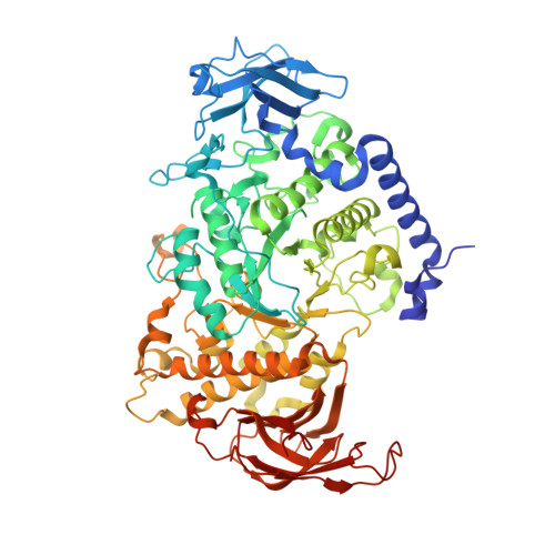
3VU2

Structure of the Starch Branching Enzyme I (BEI) complexed with maltopentaose from Oryza sativa L


Experimental Data Snapshot

  • Method: X-RAY DIFFRACTION
  • Resolution: 2.23 Å
  • R-Value Free: 0.286 
  • R-Value Work: 0.226 
  • R-Value Observed: 0.229 

wwPDB Validation   3D Report Full Report


Ligand Structure Quality Assessment 


This is version 2.1 of the entry. See complete history


Literature

Crystal structure of the rice branching enzyme I (BEI) in complex with maltopentaose.

Chaen, K.Noguchi, J.Omori, T.Kakuta, Y.Kimura, M.

(2012) Biochem Biophys Res Commun 424: 508-511

  • DOI: https://doi.org/10.1016/j.bbrc.2012.06.145
  • Primary Citation of Related Structures:  
    3VU2

  • PubMed Abstract: 

    Starch branching enzyme (SBE) catalyzes the cleavage of α-1,4-linkages and the subsequent transfer of α-1,4 glucan to form an α-1,6 branch point in amylopectin. We determined the crystal structure of the rice branching enzyme I (BEI) in complex with maltopentaose at a resolution of 2.2Å. Maltopentaose bound to a hydrophobic pocket formed by the N-terminal helix, carbohydrate-binding module 48 (CBM48), and α-amylase domain. In addition, glucose moieties could be observed at molecular surfaces on the N-terminal helix (α2) and CBM48. Amino acid residues involved in the carbohydrate bindings are highly conserved in other SBEs, suggesting their generally conserved role in substrate binding for SBEs.


  • Organizational Affiliation

    Laboratory of Structural Biology, Graduate School of Systems Life Sciences, Kyushu University, Hakozaki 6-10-1, Fukuoka 812-8581, Japan.


Macromolecules
Find similar proteins by:  (by identity cutoff)  |  3D Structure
Entity ID: 1
MoleculeChains Sequence LengthOrganismDetailsImage
1,4-alpha-glucan-branching enzyme, chloroplastic/amyloplastic
A, B
702Oryza sativa Japonica GroupMutation(s): 1 
Gene Names: SBE1RBE1Os06g0726400LOC_Os06g51084P0017G10.8-1P0017G10.8-2P0548E04.28-1P0548E04.28-2
EC: 2.4.1.18
UniProt
Find proteins for Q01401 (Oryza sativa subsp. japonica)
Explore Q01401 
Go to UniProtKB:  Q01401
Entity Groups  
Sequence Clusters30% Identity50% Identity70% Identity90% Identity95% Identity100% Identity
UniProt GroupQ01401
Sequence Annotations
Expand
  • Reference Sequence
Oligosaccharides

Help

Entity ID: 2
MoleculeChains Length2D Diagram Glycosylation3D Interactions
alpha-D-glucopyranose-(1-4)-alpha-D-glucopyranose-(1-4)-alpha-D-glucopyranose-(1-4)-alpha-D-glucopyranose-(1-4)-alpha-D-glucopyranose
C, D
5N/A
Glycosylation Resources
GlyTouCan:  G50146AM
GlyCosmos:  G50146AM
Biologically Interesting Molecules (External Reference) 1 Unique
Experimental Data & Validation

Experimental Data

  • Method: X-RAY DIFFRACTION
  • Resolution: 2.23 Å
  • R-Value Free: 0.286 
  • R-Value Work: 0.226 
  • R-Value Observed: 0.229 
  • Space Group: P 1 21 1
Unit Cell:
Length ( Å )Angle ( ˚ )
a = 73.81α = 90
b = 66.56β = 89.97
c = 169.625γ = 90
Software Package:
Software NamePurpose
ADSCdata collection
MOLREPphasing
REFMACrefinement
HKL-2000data reduction
HKL-2000data scaling

Structure Validation

View Full Validation Report


Ligand Structure Quality Assessment 


Entry History 

Deposition Data

Revision History  (Full details and data files)

  • Version 1.0: 2013-05-08
    Type: Initial release
  • Version 2.0: 2020-07-29
    Type: Remediation
    Reason: Carbohydrate remediation
    Changes: Atomic model, Data collection, Database references, Derived calculations, Non-polymer description, Structure summary
  • Version 2.1: 2024-03-20
    Changes: Data collection, Database references, Structure summary