3VLV

Crystal structure of Sphingomonas sp. A1 alginate-binding ptotein AlgQ1 in complex with unsaturated triguluronate


Experimental Data Snapshot

  • Method: X-RAY DIFFRACTION
  • Resolution: 1.50 Å
  • R-Value Free: 0.204 
  • R-Value Work: 0.185 
  • R-Value Observed: 0.186 

wwPDB Validation   3D Report Full Report


This is version 2.1 of the entry. See complete history


Literature

Recognition of heteropolysaccharide alginate by periplasmic solute-binding proteins of a bacterial ABC transporter

Nishitani, Y.Maruyama, Y.Itoh, T.Mikami, B.Hashimoto, W.Murata, K.

(2012) Biochemistry 51: 3622-3633

  • DOI: https://doi.org/10.1021/bi300194f
  • Primary Citation of Related Structures:  
    3VLU, 3VLV, 3VLW

  • PubMed Abstract: 

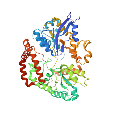
    Alginate is a heteropolysaccharide that consists of β-D-mannuronate (M) and α-L-guluronate (G). The Gram-negative bacterium Sphingomonas sp. A1 directly incorporates alginate into the cytoplasm through the periplasmic solute-binding protein (AlgQ1 and AlgQ2)-dependent ABC transporter (AlgM1-AlgM2/AlgS-AlgS). Two binding proteins with at least four subsites strongly recognize the nonreducing terminal residue of alginate at subsite 1. Here, we show the broad substrate preference of strain A1 solute-binding proteins for M and G present in alginate and demonstrate the structural determinants in binding proteins for heteropolysaccharide recognition through X-ray crystallography of four AlgQ1 structures in complex with saturated and unsaturated alginate oligosaccharides. Alginates with different M/G ratios were assimilated by strain A1 cells and bound to AlgQ1 and AlgQ2. Crystal structures of oligosaccharide-bound forms revealed that in addition to interaction between AlgQ1 and unsaturated oligosaccharides, the binding protein binds through hydrogen bonds to the C4 hydroxyl group of the saturated nonreducing terminal residue at subsite 1. The M residue of saturated oligosaccharides is predominantly accommodated at subsite 1 because of the strict binding of Ser-273 to the carboxyl group of the residue. In unsaturated trisaccharide (ΔGGG or ΔMMM)-bound AlgQ1, the protein interacts appropriately with substrate hydroxyl groups at subsites 2 and 3 to accommodate M or G, while substrate carboxyl groups are strictly recognized by the specific residues Tyr-129 at subsite 2 and Lys-22 at subsite 3. Because of this substrate recognition mechanism, strain A1 solute-binding proteins can bind heteropolysaccharide alginate with different M/G ratios.


  • Organizational Affiliation

    Laboratory of Basic and Applied Molecular Biotechnology, Graduate School of Agriculture, Kyoto University, Uji, Kyoto 611-0011, Japan.


Macromolecules
Find similar proteins by:  (by identity cutoff)  |  3D Structure
Entity ID: 1
MoleculeChains Sequence LengthOrganismDetailsImage
AlgQ1502Sphingomonas sp. A1Mutation(s): 0 
Gene Names: algQ1
UniProt
Find proteins for Q9KWT6 (Sphingomonas sp)
Explore Q9KWT6 
Go to UniProtKB:  Q9KWT6
Entity Groups  
Sequence Clusters30% Identity50% Identity70% Identity90% Identity95% Identity100% Identity
UniProt GroupQ9KWT6
Sequence Annotations
Expand
  • Reference Sequence
Oligosaccharides

Help

Entity ID: 2
MoleculeChains Length2D Diagram Glycosylation3D Interactions
4-deoxy-alpha-L-erythro-hex-4-enopyranuronic acid-(1-4)-alpha-L-gulopyranuronic acid-(1-4)-alpha-L-gulopyranuronic acid
B
3N/A
Glycosylation Resources
GlyTouCan:  G31814BT
GlyCosmos:  G31814BT
Small Molecules
Ligands 1 Unique
IDChains Name / Formula / InChI Key2D Diagram3D Interactions
CA
Query on CA

Download Ideal Coordinates CCD File 
C [auth A]CALCIUM ION
Ca
BHPQYMZQTOCNFJ-UHFFFAOYSA-N
Experimental Data & Validation

Experimental Data

  • Method: X-RAY DIFFRACTION
  • Resolution: 1.50 Å
  • R-Value Free: 0.204 
  • R-Value Work: 0.185 
  • R-Value Observed: 0.186 
  • Space Group: P 1 21 1
Unit Cell:
Length ( Å )Angle ( ˚ )
a = 58.369α = 90
b = 67.517β = 94.57
c = 61.558γ = 90
Software Package:
Software NamePurpose
MOLREPphasing
REFMACrefinement
HKL-2000data reduction
HKL-2000data scaling

Structure Validation

View Full Validation Report


Entry History 

Deposition Data

Revision History  (Full details and data files)

  • Version 1.0: 2012-01-25
    Type: Initial release
  • Version 1.1: 2013-11-27
    Changes: Database references
  • Version 2.0: 2020-07-29
    Type: Remediation
    Reason: Carbohydrate remediation
    Changes: Atomic model, Data collection, Derived calculations, Structure summary
  • Version 2.1: 2023-11-08
    Changes: Data collection, Database references, Refinement description, Structure summary