


Crystal Structure of VldE, the pseudo-glycosyltransferase which catalyzes non-glycosidic C-N coupling in Validamycin A biosynthesis
Cavalier, M.C., Yim, Y.-S., Asamizu, S., Neau, D., Mahmud, T., Lee, Y.-H.To be published.
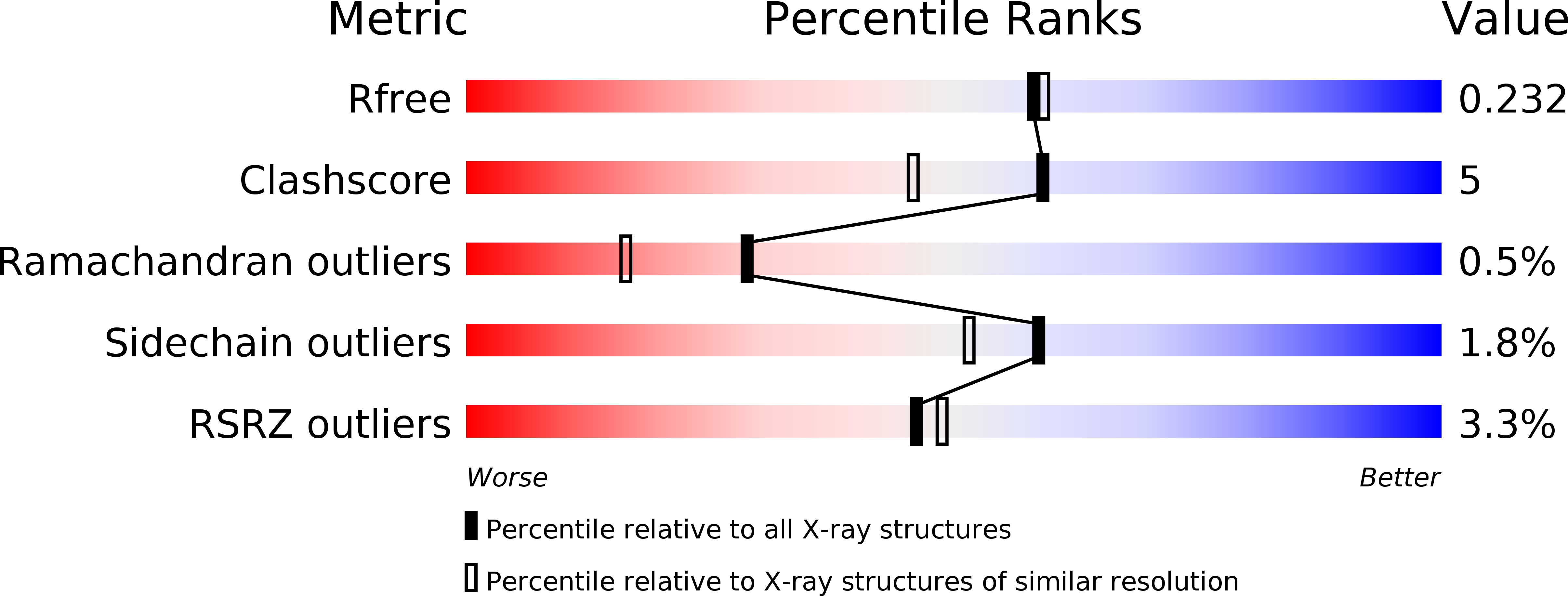
Experimental Data Snapshot
wwPDB Validation 3D Report Full Report
Entity ID: 1 | |||||
|---|---|---|---|---|---|
| Molecule | Chains | Sequence Length | Organism | Details | Image |
| VldE | 497 | Streptomyces hygroscopicus subsp. limoneus | Mutation(s): 0 Gene Names: vldE EC: 2.4 | 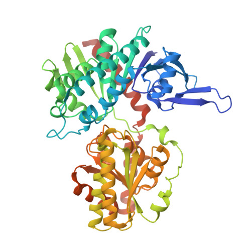 | |
UniProt | |||||
Find proteins for Q15JG1 (Streptomyces hygroscopicus subsp. limoneus) Explore Q15JG1 Go to UniProtKB: Q15JG1 | |||||
Entity Groups | |||||
| Sequence Clusters | 30% Identity50% Identity70% Identity90% Identity95% Identity100% Identity | ||||
| UniProt Group | Q15JG1 | ||||
Sequence AnnotationsExpand | |||||
| |||||
| Length ( Å ) | Angle ( ˚ ) |
|---|---|
| a = 85.237 | α = 90 |
| b = 48.562 | β = 91.88 |
| c = 123.049 | γ = 90 |
| Software Name | Purpose |
|---|---|
| ADSC | data collection |
| PHENIX | model building |
| REFMAC | refinement |
| XDS | data reduction |
| XDS | data scaling |
| PHENIX | phasing |
RCSB PDB (citation) is hosted by
RCSB PDB is a member of the

