3V8C

Crystal structure of monoclonal human anti-rhesus D Fc IgG1 t125(yb2/0) double mutant (H310 and H435 in K)


Experimental Data Snapshot

  • Method: X-RAY DIFFRACTION
  • Resolution: 2.77 Å
  • R-Value Free: 0.316 
  • R-Value Work: 0.254 
  • R-Value Observed: 0.258 

wwPDB Validation   3D Report Full Report


Ligand Structure Quality Assessment 


This is version 2.1 of the entry. See complete history


Literature

Effect of zinc on human IgG1 and its Fc gamma R interactions.

Siberil, S.Menez, R.Jorieux, S.de Romeuf, C.Bourel, D.Fridman, W.H.Ducancel, F.Stura, E.A.Teillaud, J.L.

(2012) Immunol Lett 143: 60-69

  • DOI: https://doi.org/10.1016/j.imlet.2012.02.002
  • Primary Citation of Related Structures:  
    3V7M, 3V8C, 3V95

  • PubMed Abstract: 

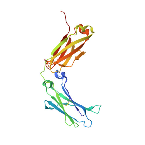
    In the present study, we show that histidines 310 and 435 at the CH2-CH3 interface of the Fc portion of human IgG1 can coordinate a Zn2+ and participate in the control of the CH2-CH2 interdomain opening. Structures obtained in the absence of Zn2+ have a reduced interdomain gap that likely hamper FcγR binding. This closed conformation of the Fc is stabilized by inter-CH2 domain sugar contacts. Zinc appears to counteract the sugar mediated constriction, suggesting that zinc could be an important control factor in IgG1/FcγR interactions. The results of binding studies performed in the presence of EDTA on FcγR expressing cells supports this hypothesis. When a mutated Fc fragment, in which histidines 310 and 435 have been substituted by lysines (Fc H/K), was compared with the wild-type Fc in crystallographic studies, we found that the mutations leave the interface unaltered but have a long-range effect on the CH2 interdomain separation. Moreover, these substitutions have a differential effect on the binding of IgG1 to Fcγ receptors and their functions. Interaction with the inhibitory FcγRIIB is strongly perturbed by the mutations and mutant IgG1 H/K only weakly engages this receptor. By contrast, higher affinity FcγR are mostly unaffected.


  • Organizational Affiliation

    INSERM UMR S 872, Centre de Recherche des Cordeliers, Paris, France.


Macromolecules
Find similar proteins by:  (by identity cutoff)  |  3D Structure
Entity ID: 1
MoleculeChains Sequence LengthOrganismDetailsImage
Ig gamma-1 chain C region
A, B
212Homo sapiensMutation(s): 2 
Gene Names: HUMANIGHG1
UniProt & NIH Common Fund Data Resources
Find proteins for P01857 (Homo sapiens)
Explore P01857 
Go to UniProtKB:  P01857
PHAROS:  P01857
GTEx:  ENSG00000211896 
Entity Groups  
Sequence Clusters30% Identity50% Identity70% Identity90% Identity95% Identity100% Identity
UniProt GroupP01857
Sequence Annotations
Expand
  • Reference Sequence
Oligosaccharides

Help

Entity ID: 2
MoleculeChains Length2D Diagram Glycosylation3D Interactions
2-acetamido-2-deoxy-beta-D-glucopyranose-(1-2)-alpha-D-mannopyranose
C
2N/A
Glycosylation Resources
GlyTouCan:  G85856KC
GlyCosmos:  G85856KC
GlyGen:  G85856KC
Small Molecules
Ligands 4 Unique
IDChains Name / Formula / InChI Key2D Diagram3D Interactions
NAG
Query on NAG

Download Ideal Coordinates CCD File 
D [auth A]
E [auth A]
J [auth A]
M [auth A]
N [auth B]
D [auth A],
E [auth A],
J [auth A],
M [auth A],
N [auth B],
P [auth B],
S [auth B]
2-acetamido-2-deoxy-beta-D-glucopyranose
C8 H15 N O6
OVRNDRQMDRJTHS-FMDGEEDCSA-N
GAL
Query on GAL

Download Ideal Coordinates CCD File 
K [auth A],
T [auth B]
beta-D-galactopyranose
C6 H12 O6
WQZGKKKJIJFFOK-FPRJBGLDSA-N
MAN
Query on MAN

Download Ideal Coordinates CCD File 
G [auth A],
I [auth A],
L [auth A],
Q [auth B],
R [auth B]
alpha-D-mannopyranose
C6 H12 O6
WQZGKKKJIJFFOK-PQMKYFCFSA-N
GOL
Query on GOL

Download Ideal Coordinates CCD File 
F [auth A],
H [auth A],
O [auth B]
GLYCEROL
C3 H8 O3
PEDCQBHIVMGVHV-UHFFFAOYSA-N
Experimental Data & Validation

Experimental Data

  • Method: X-RAY DIFFRACTION
  • Resolution: 2.77 Å
  • R-Value Free: 0.316 
  • R-Value Work: 0.254 
  • R-Value Observed: 0.258 
  • Space Group: P 21 21 21
Unit Cell:
Length ( Å )Angle ( ˚ )
a = 49.54α = 90
b = 79.4β = 90
c = 139.65γ = 90
Software Package:
Software NamePurpose
DNAdata collection
MOLREPphasing
REFMACrefinement
XDSdata reduction
SCALAdata scaling

Structure Validation

View Full Validation Report


Ligand Structure Quality Assessment 


Entry History 

Deposition Data

Revision History  (Full details and data files)

  • Version 1.0: 2012-02-22
    Type: Initial release
  • Version 1.1: 2013-08-28
    Changes: Database references
  • Version 1.2: 2014-10-08
    Changes: Structure summary
  • Version 2.0: 2020-07-29
    Type: Remediation
    Reason: Carbohydrate remediation
    Changes: Advisory, Atomic model, Data collection, Database references, Derived calculations, Structure summary
  • Version 2.1: 2023-09-13
    Changes: Data collection, Database references, Refinement description, Structure summary