3V7T

Crystal Structure of Human Beta-Tryptase Complexed with a Synthetic Inhibitor with a Tropanylamide Scaffold


Experimental Data Snapshot

  • Method: X-RAY DIFFRACTION
  • Resolution: 2.09 Å
  • R-Value Free: 0.234 
  • R-Value Work: 0.208 
  • R-Value Observed: 0.210 

wwPDB Validation   3D Report Full Report


Ligand Structure Quality Assessment 


This is version 1.1 of the entry. See complete history


Literature

A beta-tryptase inhibitor with a tropanylamide scaffold to improve in vitro stability and to lower hERG channel binding affinity

Liang, G.Choi-Sledeski, Y.M.Shum, P.Chen, X.Poli, G.B.Kumar, V.Minnich, A.Wang, Q.Tsay, J.Sides, K.Kang, J.Zhang, Y.

(2012) Bioorg Med Chem Lett 22: 1606-1610

  • DOI: https://doi.org/10.1016/j.bmcl.2011.12.127
  • Primary Citation of Related Structures:  
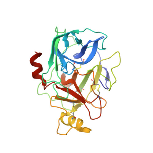
    3V7T

  • PubMed Abstract: 

    Tropanylamide was investigated as a possible scaffold for β-tryptase inhibitors with a basic benzylamine P1 group and a substituted thiophene P4 group. Comparing to piperidinylamide, the tropanylamide scaffold is much more rigid, which presents less opportunity for the inhibitor to bind with off-target proteins, such as cytochrome P450, SSAO, and hERG potassium channel. The proposed binding mode was further confirmed by an in-house X-ray structure through co-crystallization.


  • Organizational Affiliation

    Molecular Innovative Therapeutics, Sanofi Pharmaceuticals, Bridgewater, NJ 08807, USA. guyan.liang@sanofi-aventis.com


Macromolecules
Find similar proteins by:  (by identity cutoff)  |  3D Structure
Entity ID: 1
MoleculeChains Sequence LengthOrganismDetailsImage
TPSB2 protein
A, B, C, D
245Homo sapiensMutation(s): 0 
Gene Names: TPSB2
UniProt & NIH Common Fund Data Resources
Find proteins for P20231 (Homo sapiens)
Explore P20231 
Go to UniProtKB:  P20231
PHAROS:  P20231
GTEx:  ENSG00000197253 
Entity Groups  
Sequence Clusters30% Identity50% Identity70% Identity90% Identity95% Identity100% Identity
UniProt GroupP20231
Sequence Annotations
Expand
  • Reference Sequence
Small Molecules
Binding Affinity Annotations 
IDSourceBinding Affinity
0GX PDBBind:  3V7T Ki: 62 (nM) from 1 assay(s)
Binding MOAD:  3V7T Ki: 62 (nM) from 1 assay(s)
Experimental Data & Validation

Experimental Data

  • Method: X-RAY DIFFRACTION
  • Resolution: 2.09 Å
  • R-Value Free: 0.234 
  • R-Value Work: 0.208 
  • R-Value Observed: 0.210 
  • Space Group: P 41
Unit Cell:
Length ( Å )Angle ( ˚ )
a = 81.719α = 90
b = 81.719β = 90
c = 170.684γ = 90
Software Package:
Software NamePurpose
DENZOdata reduction
SCALEPACKdata scaling
MOLREPphasing
BUSTER-TNTrefinement
PDB_EXTRACTdata extraction
HKL-2000data reduction
HKL-2000data scaling
BUSTERrefinement

Structure Validation

View Full Validation Report


Ligand Structure Quality Assessment 


Entry History 

Deposition Data

Revision History  (Full details and data files)

  • Version 1.0: 2012-03-14
    Type: Initial release
  • Version 1.1: 2017-11-08
    Changes: Refinement description