3UIY

Crystal structure of SefD_dscA in H2O


Experimental Data Snapshot

  • Method: X-RAY DIFFRACTION
  • Resolution: 3.10 Å
  • R-Value Free: 0.332 
  • R-Value Work: 0.268 
  • R-Value Observed: 0.270 

wwPDB Validation   3D Report Full Report


This is version 1.4 of the entry. See complete history


Literature

Promoting crystallisation of the Salmonella enteritidis fimbriae 14 pilin SefD using deuterium oxide.

Liu, B.Garnett, J.A.Lee, W.C.Lin, J.Salgado, P.Taylor, J.Xu, Y.Lambert, S.Cota, E.Matthews, S.

(2012) Biochem Biophys Res Commun 421: 208-213

  • DOI: https://doi.org/10.1016/j.bbrc.2012.03.136
  • Primary Citation of Related Structures:  
    3UIY, 3UIZ

  • PubMed Abstract: 

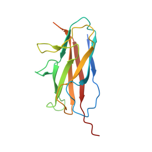
    The use of heavy water (D(2)O) as a solvent is commonplace in many spectroscopic techniques for the study of biological macromolecules. A significant deuterium isotope effect exists where hydrogen-bonding is important, such as in protein stability, dynamics and assembly. Here we illustrate the use of D(2)O in additive screening for the production of reproducible diffraction-quality crystals for the Salmonella enteritidis fimbriae 14 (SEF14) putative tip adhesin, SefD.


  • Organizational Affiliation

    Centre for Structural Biology & Division of Molecular Biosciences, Imperial College London, South Kensington, London SW7 2AZ, UK.


Macromolecules
Find similar proteins by:  (by identity cutoff)  |  3D Structure
Entity ID: 1
MoleculeChains Sequence LengthOrganismDetailsImage
Chimera protein of SefD and SefA152Salmonella enterica subsp. enterica serovar EnteritidisMutation(s): 0 
Gene Names: sefD
UniProt
Find proteins for Q53997 (Salmonella enteritidis)
Explore Q53997 
Go to UniProtKB:  Q53997
Find proteins for P12061 (Salmonella enteritidis)
Explore P12061 
Go to UniProtKB:  P12061
Entity Groups  
Sequence Clusters30% Identity50% Identity70% Identity90% Identity95% Identity100% Identity
UniProt GroupsQ53997P12061
Sequence Annotations
Expand
  • Reference Sequence
Experimental Data & Validation

Experimental Data

  • Method: X-RAY DIFFRACTION
  • Resolution: 3.10 Å
  • R-Value Free: 0.332 
  • R-Value Work: 0.268 
  • R-Value Observed: 0.270 
  • Space Group: P 65 2 2
Unit Cell:
Length ( Å )Angle ( ˚ )
a = 52.48α = 90
b = 52.48β = 90
c = 218.03γ = 120
Software Package:
Software NamePurpose
Adxvdata processing
PHASERphasing
REFMACrefinement
xia2data reduction
SCALAdata scaling

Structure Validation

View Full Validation Report


Entry History 

Deposition Data

Revision History  (Full details and data files)

  • Version 1.0: 2012-05-02
    Type: Initial release
  • Version 1.1: 2013-01-09
    Changes: Database references
  • Version 1.2: 2017-07-26
    Changes: Data collection, Refinement description, Source and taxonomy
  • Version 1.3: 2017-11-08
    Changes: Refinement description
  • Version 1.4: 2023-09-13
    Changes: Data collection, Database references, Refinement description