3TVZ

Structure of Bacillus subtilis HmoB


Experimental Data Snapshot

  • Method: X-RAY DIFFRACTION
  • Resolution: 2.00 Å
  • R-Value Free: 0.251 
  • R-Value Work: 0.196 
  • R-Value Observed: 0.199 

wwPDB Validation   3D Report Full Report


This is version 1.3 of the entry. See complete history


Literature

Bacillus subtilis HmoB is a heme oxygenase with a novel structure.

Park, S.Choi, S.Choe, J.

(2012) BMB Rep 45: 239-241

  • DOI: https://doi.org/10.5483/bmbrep.2012.45.4.239
  • Primary Citation of Related Structures:  
    3TVZ

  • PubMed Abstract: 

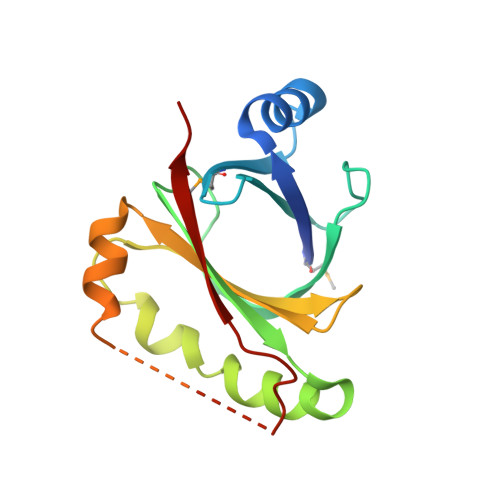
    Iron availability is limited in the environment and most bacteria have developed a system to acquire iron from host hemoproteins. Heme oxygenase plays an important role by degrading heme group and releasing the essential nutrient iron. The structure of Bacillus subtilis HmoB was determined to 2.0 A resolution. B. subtilis HmoB contains a typical antibiotic biosynthesis monooxygenase (ABM) domain that spans from 71 to 146 residues and belongs to the IsdG family heme oxygenases. Comparison of HmoB and IsdG family proteins showed that the C-terminal region of HmoB has similar sequence and structure to IsdG family proteins and contains conserved critical residues for heme degradation. However, HmoB is distinct from other IsdG family proteins in that HmoB is about 60 amino acids longer in the N-terminus and does not form a dimer whereas previously studied IsdG family heme oxygenases form functional homodimers. Interestingly, the structure of monomeric HmoB resembles the dimeric structure of IsdG family proteins. Hence, B. subtilis HmoB is a heme oxygenase with a novel structural feature.


  • Organizational Affiliation

    Department of Life Science, University of Seoul, Seoul, Korea.


Macromolecules
Find similar proteins by:  (by identity cutoff)  |  3D Structure
Entity ID: 1
MoleculeChains Sequence LengthOrganismDetailsImage
Putative uncharacterized protein yhgC
A, B, C
172Bacillus spizizenii str. W23Mutation(s): 0 
Gene Names: yhgCBSUW23_05110
UniProt
Find proteins for E0TXX3 (Bacillus spizizenii (strain ATCC 23059 / NRRL B-14472 / W23))
Explore E0TXX3 
Go to UniProtKB:  E0TXX3
Entity Groups  
Sequence Clusters30% Identity50% Identity70% Identity90% Identity95% Identity100% Identity
UniProt GroupE0TXX3
Sequence Annotations
Expand
  • Reference Sequence
Small Molecules
Modified Residues  1 Unique
IDChains TypeFormula2D DiagramParent
MSE
Query on MSE
A, B, C
L-PEPTIDE LINKINGC5 H11 N O2 SeMET
Experimental Data & Validation

Experimental Data

  • Method: X-RAY DIFFRACTION
  • Resolution: 2.00 Å
  • R-Value Free: 0.251 
  • R-Value Work: 0.196 
  • R-Value Observed: 0.199 
  • Space Group: P 61 2 2
Unit Cell:
Length ( Å )Angle ( ˚ )
a = 64.799α = 90
b = 64.799β = 90
c = 392.591γ = 120
Software Package:
Software NamePurpose
DENZOdata reduction
SCALEPACKdata scaling
REFMACrefinement
PDB_EXTRACTdata extraction
HKL-2000data collection
SOLVEphasing

Structure Validation

View Full Validation Report


Entry History 

Deposition Data

Revision History  (Full details and data files)

  • Version 1.0: 2012-07-11
    Type: Initial release
  • Version 1.1: 2012-07-18
    Changes: Structure summary
  • Version 1.2: 2017-11-08
    Changes: Refinement description
  • Version 1.3: 2019-10-09
    Changes: Data collection, Database references, Derived calculations