3TVI

Crystal structure of Clostridium acetobutylicum aspartate kinase (CaAK): An important allosteric enzyme for industrial amino acids production


Experimental Data Snapshot

  • Method: X-RAY DIFFRACTION
  • Resolution: 3.00 Å
  • R-Value Free: 0.273 
  • R-Value Work: 0.206 
  • R-Value Observed: 0.207 

wwPDB Validation   3D Report Full Report


This is version 1.2 of the entry. See complete history


Literature

Crystal structure of Clostridium acetobutylicum Aspartate kinase (CaAK): An important allosteric enzyme for amino acids production.

Manjasetty, B.A.Chance, M.R.Burley, S.K.Panjikar, S.Almo, S.C.

(2014) Biotechnol Rep (amst) 3: 73-85

  • DOI: https://doi.org/10.1016/j.btre.2014.06.009
  • Primary Citation of Related Structures:  
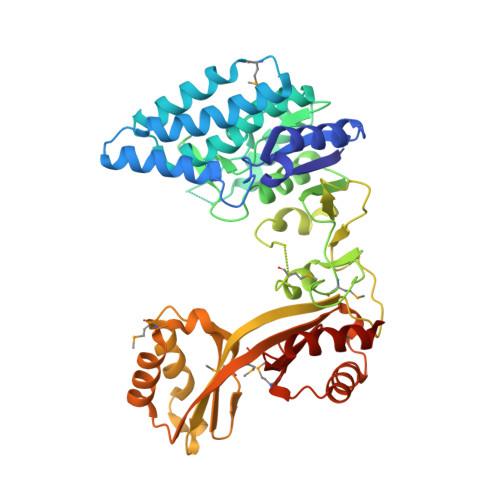
    3TVI

  • PubMed Abstract: 

    Aspartate kinase (AK) is an enzyme which is tightly regulated through feedback control and responsible for the synthesis of 4-phospho-L-aspartate from L-aspartate. This intermediate step is at an important branch point where one path leads to the synthesis of lysine and the other to threonine, methionine and isoleucine. Concerted feedback inhibition of AK is mediated by threonine and lysine and varies between the species. The crystal structure of biotechnologically important Clostridium acetobutylicum aspartate kinase ( Ca AK; E.C. 2.7.2.4; Mw =48,030Da; 437aa; SwissProt: Q97MC0) has been determined to 3Å resolution. Ca AK acquires a protein fold similar to the other known structures of AKs despite the low sequence identity (<30%). It is composed of two domains: an N-terminal catalytic domain (kinase) domain and a C-terminal regulatory domain further comprised of two small domains belonging to the ACT domain family. Pairwise comparison of 12 molecules in the asymmetric unit helped to identify the bending regions which are in the vicinity of ATP binding site involved in domain movements between the catalytic and regulatory domains. All 12 Ca AK molecules adopt fully open T-state conformation leading to the formation of three tetramers unique among other similar AK structures. On the basis of comparative structural analysis, we discuss tetramer formation based on the large conformational changes in the catalytic domain associated with the lysine binding at the regulatory domains. The structure described herein is homologous to a target in wide-spread pathogenic (toxin producing) bacteria such as Clostridium tetani (64% sequence identity) suggesting the potential of the structure solved here to be applied for modeling drug interactions. Ca AK structure may serve as a guide to better understand and engineer lysine biosynthesis for the biotechnology industry.


Macromolecules
Find similar proteins by:  (by identity cutoff)  |  3D Structure
Entity ID: 1
MoleculeChains Sequence LengthOrganismDetailsImage
Aspartokinase
A, B, C, D, E
A, B, C, D, E, F, G, H, I, J, K, L
446Clostridium acetobutylicumMutation(s): 0 
Gene Names: CA_C0278
EC: 2.7.2.4
UniProt
Find proteins for Q97MC0 (Clostridium acetobutylicum (strain ATCC 824 / DSM 792 / JCM 1419 / LMG 5710 / VKM B-1787))
Explore Q97MC0 
Go to UniProtKB:  Q97MC0
Entity Groups  
Sequence Clusters30% Identity50% Identity70% Identity90% Identity95% Identity100% Identity
UniProt GroupQ97MC0
Sequence Annotations
Expand
  • Reference Sequence
Small Molecules
Ligands 2 Unique
IDChains Name / Formula / InChI Key2D Diagram3D Interactions
LYS
Query on LYS

Download Ideal Coordinates CCD File 
N [auth A]
O [auth B]
P [auth C]
R [auth D]
U [auth G]
N [auth A],
O [auth B],
P [auth C],
R [auth D],
U [auth G],
W [auth H]
LYSINE
C6 H15 N2 O2
KDXKERNSBIXSRK-YFKPBYRVSA-O
ASP
Query on ASP

Download Ideal Coordinates CCD File 
M [auth A]
Q [auth D]
S [auth E]
T [auth G]
V [auth H]
M [auth A],
Q [auth D],
S [auth E],
T [auth G],
V [auth H],
X [auth I],
Y [auth L]
ASPARTIC ACID
C4 H7 N O4
CKLJMWTZIZZHCS-REOHCLBHSA-N
Modified Residues  1 Unique
IDChains TypeFormula2D DiagramParent
MSE
Query on MSE
A, B, C, D, E
A, B, C, D, E, F, G, H, I, J, K, L
L-PEPTIDE LINKINGC5 H11 N O2 SeMET
Experimental Data & Validation

Experimental Data

  • Method: X-RAY DIFFRACTION
  • Resolution: 3.00 Å
  • R-Value Free: 0.273 
  • R-Value Work: 0.206 
  • R-Value Observed: 0.207 
  • Space Group: P 1 21 1
Unit Cell:
Length ( Å )Angle ( ˚ )
a = 109.041α = 90
b = 274.217β = 113.69
c = 114.041γ = 90
Software Package:
Software NamePurpose
CBASSdata collection
SHELXDphasing
REFMACrefinement
HKL-2000data reduction
HKL-2000data scaling

Structure Validation

View Full Validation Report


Entry History 

Revision History  (Full details and data files)

  • Version 1.0: 2011-11-09
    Type: Initial release
  • Version 1.1: 2014-09-17
    Changes: Database references
  • Version 1.2: 2021-02-10
    Changes: Database references, Derived calculations, Structure summary