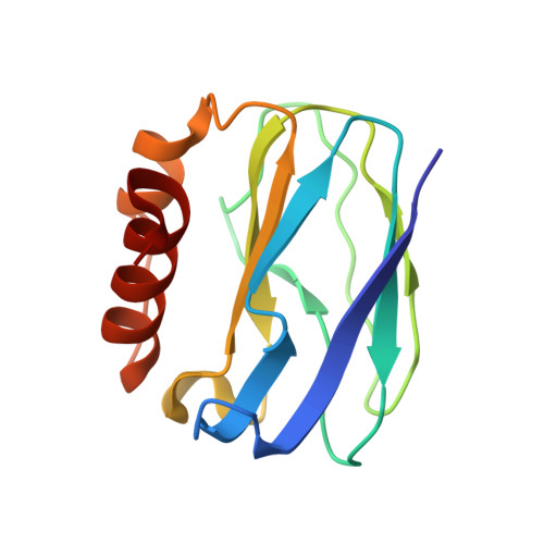
3TU6

The Structure of a Pseudoazurin From Sinorhizobium meliltoi


Experimental Data Snapshot

  • Method: X-RAY DIFFRACTION
  • Resolution: 2.00 Å
  • R-Value Free: 0.261 
  • R-Value Work: 0.202 
  • R-Value Observed: 0.207 

wwPDB Validation   3D Report Full Report


This is version 1.3 of the entry. See complete history


Literature

The X-ray crystal structure of a pseudoazurin from Sinorhizobium meliloti.

Laming, E.M.McGrath, A.P.Guss, J.M.Kappler, U.Maher, M.J.

(2012) J Inorg Biochem 115: 148-154

  • DOI: https://doi.org/10.1016/j.jinorgbio.2012.04.005
  • Primary Citation of Related Structures:  
    3TU6

  • PubMed Abstract: 

    The X-ray crystal structure of oxidised pseudoazurin from the denitrifying plant symbiotic bacterium Sinorhizobium meliloti (SmPAz2) has been solved to a resolution of 2.0 Å. The pseudoazurin from Sinorhizobium sp. is unusual as it forms an operon with a sulfite dehydrogenase enzyme, rather than a Cu nitrite reductase. Examination of the structure reveals that the geometric parameters of the Type I Cu site in SmPAz2 correlate with observed features in the electronic spectrum of the protein. Comparison of the structure of SmPAz2 with those of pseudoazurins from five other bacterial species shows that the surface of SmPAz2 bears a conserved hydrophobic patch encircled by positively-charged residues, which may serve as a recognition site for its redox partners.


  • Organizational Affiliation

    School of Molecular Bioscience, University of Sydney, NSW 2006, Australia.


Macromolecules
Find similar proteins by:  (by identity cutoff)  |  3D Structure
Entity ID: 1
MoleculeChains Sequence LengthOrganismDetailsImage
Pseudoazurin (Blue copper protein)127Sinorhizobium melilotiMutation(s): 0 
Gene Names: azu2AZU2TR02830SMc04047
UniProt
Find proteins for Q92M26 (Rhizobium meliloti (strain 1021))
Explore Q92M26 
Go to UniProtKB:  Q92M26
Entity Groups  
Sequence Clusters30% Identity50% Identity70% Identity90% Identity95% Identity100% Identity
UniProt GroupQ92M26
Sequence Annotations
Expand
  • Reference Sequence
Small Molecules
Experimental Data & Validation

Experimental Data

  • Method: X-RAY DIFFRACTION
  • Resolution: 2.00 Å
  • R-Value Free: 0.261 
  • R-Value Work: 0.202 
  • R-Value Observed: 0.207 
  • Space Group: P 21 21 21
Unit Cell:
Length ( Å )Angle ( ˚ )
a = 35.627α = 90
b = 53.494β = 90
c = 59.921γ = 90
Software Package:
Software NamePurpose
SCALEPACKdata scaling
REFMACrefinement
PDB_EXTRACTdata extraction
PHASERphasing

Structure Validation

View Full Validation Report


Entry History 

Deposition Data

Revision History  (Full details and data files)

  • Version 1.0: 2012-05-30
    Type: Initial release
  • Version 1.1: 2012-07-25
    Changes: Database references
  • Version 1.2: 2012-12-19
    Changes: Database references
  • Version 1.3: 2024-02-28
    Changes: Data collection, Database references, Derived calculations