3TSA

Spinosyn Rhamnosyltransferase SpnG


Experimental Data Snapshot

  • Method: X-RAY DIFFRACTION
  • Resolution: 1.70 Å
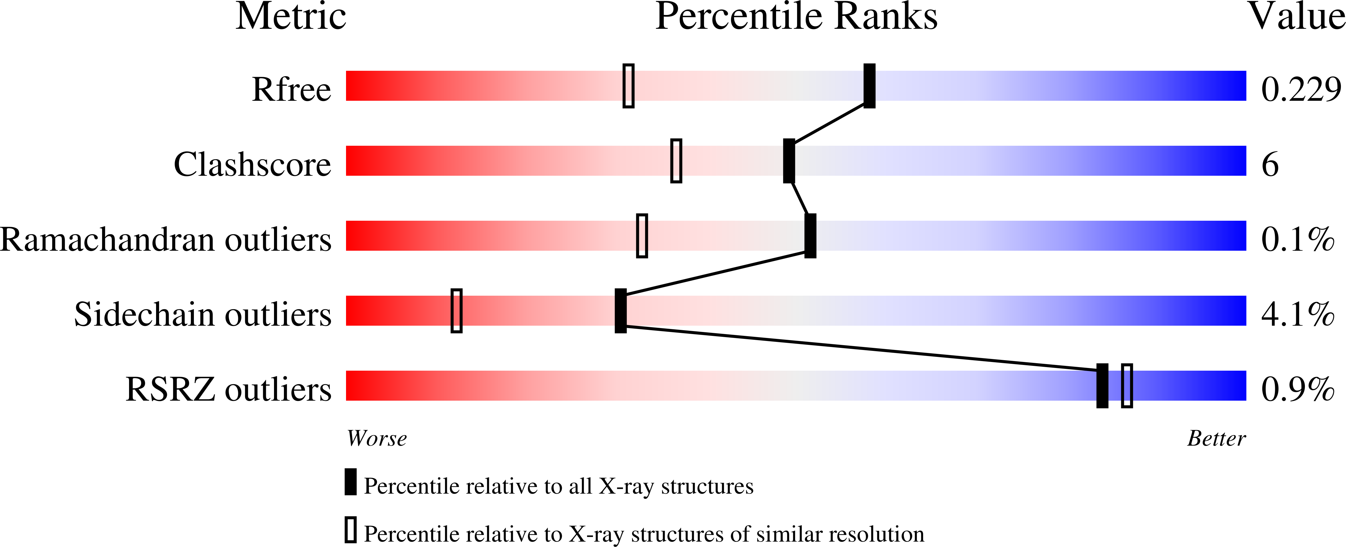
  • R-Value Free: 0.231 
  • R-Value Work: 0.181 
  • R-Value Observed: 0.184 

wwPDB Validation   3D Report Full Report


Ligand Structure Quality Assessment 


This is version 1.4 of the entry. See complete history


Literature

Structural Studies of the Spinosyn Rhamnosyltransferase, SpnG.

Isiorho, E.A.Liu, H.W.Keatinge-Clay, A.T.

(2012) Biochemistry 51: 1213-1222

  • DOI: https://doi.org/10.1021/bi201860q
  • Primary Citation of Related Structures:  
    3TSA, 3UYK, 3UYL

  • PubMed Abstract: 

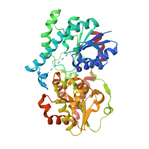
    Spinosyns A and D (spinosad), like many other complex polyketides, are tailored near the end of their biosyntheses through the addition of sugars. SpnG, which catalyzes their 9-OH rhamnosylation, is also capable of adding other monosaccharides to the spinosyn aglycone (AGL) from TDP-sugars; however, the substitution of UDP-D-glucose for TDP-D-glucose as the donor substrate is known to result in a >60000-fold reduction in k(cat). Here, we report the structure of SpnG at 1.65 Å resolution, SpnG bound to TDP at 1.86 Å resolution, and SpnG bound to AGL at 1.70 Å resolution. The SpnG-TDP complex reveals how SpnG employs N202 to discriminate between TDP- and UDP-sugars. A conformational change of several residues in the active site is promoted by the binding of TDP. The SpnG-AGL complex shows that the binding of AGL is mediated via hydrophobic interactions and that H13, the potential catalytic base, is within 3 Å of the nucleophilic 9-OH group of AGL. A model for the Michaelis complex was constructed to reveal the features that allow SpnG to transfer diverse sugars; it also revealed that the rhamnosyl moiety is in a skew-boat conformation during the transfer reaction.


  • Organizational Affiliation

    Division of Medicinal Chemistry, College of Pharmacy, The University of Texas at Austin, Austin, Texas 78712, United States.


Macromolecules
Find similar proteins by:  (by identity cutoff)  |  3D Structure
Entity ID: 1
MoleculeChains Sequence LengthOrganismDetailsImage
NDP-rhamnosyltransferase
A, B
391Saccharopolyspora spinosaMutation(s): 0 
Gene Names: spnG
EC: 2.4.1
UniProt
Find proteins for Q9ALM8 (Saccharopolyspora spinosa)
Explore Q9ALM8 
Go to UniProtKB:  Q9ALM8
Entity Groups  
Sequence Clusters30% Identity50% Identity70% Identity90% Identity95% Identity100% Identity
UniProt GroupQ9ALM8
Sequence Annotations
Expand
  • Reference Sequence
Small Molecules
Experimental Data & Validation

Experimental Data

  • Method: X-RAY DIFFRACTION
  • Resolution: 1.70 Å
  • R-Value Free: 0.231 
  • R-Value Work: 0.181 
  • R-Value Observed: 0.184 
  • Space Group: P 1
Unit Cell:
Length ( Å )Angle ( ˚ )
a = 54.16α = 81.46
b = 57.06β = 73.84
c = 68.113γ = 85.95
Software Package:
Software NamePurpose
REFMACrefinement
CNSrefinement
StructureStudiodata collection
HKL-2000data reduction
HKL-2000data scaling
CNSphasing

Structure Validation

View Full Validation Report


Ligand Structure Quality Assessment 


Entry History 

Deposition Data

Revision History  (Full details and data files)

  • Version 1.0: 2012-02-15
    Type: Initial release
  • Version 1.1: 2012-02-29
    Changes: Database references
  • Version 1.2: 2019-07-17
    Changes: Data collection, Refinement description
  • Version 1.3: 2020-07-29
    Type: Remediation
    Reason: Carbohydrate remediation
    Changes: Data collection, Database references, Derived calculations, Structure summary
  • Version 1.4: 2023-09-13
    Changes: Data collection, Database references, Refinement description, Structure summary