3THK

Structure of SH3 chimera with a type II ligand linked to the chain C-terminal


Experimental Data Snapshot

  • Method: X-RAY DIFFRACTION
  • Resolution: 1.70 Å
  • R-Value Free: 0.201 
  • R-Value Work: 0.180 
  • R-Value Observed: 0.181 

wwPDB Validation   3D Report Full Report


This is version 1.2 of the entry. See complete history


Literature

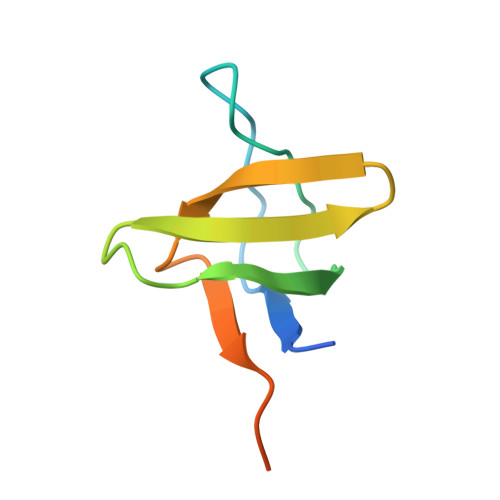
High-Resolution Crystal Structure of Spectrin SH3 Domain Fused with a Proline-Rich Peptide.

Gushchina, L.V.Gabdulkhakov, A.G.Nikonov, S.V.Filimonov, V.V.

(2011) J Biomol Struct Dyn 29: 485-495

  • DOI: https://doi.org/10.1080/07391102.2011.10507400
  • Primary Citation of Related Structures:  
    3THK

  • PubMed Abstract: 

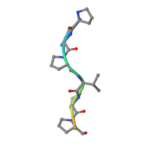
    A new chimeric protein, named WT-CIIA, was designed by connecting the proline-rich decapeptide PPPVPPYSAG to the C-terminus of the alpha-spectrin SH3 domain through a natural twelve-residue linker to obtain a single-chain model that would imitate intramolecular SH3-ligand interaction. The crystal structure of this fusion protein was determined at 1.7 Å resolution. The asymmetric unit of the crystal contained two SH3 globules contacting with one PPPVPPY fragment located between them. The domains are related by the two-fold non-crystallographic axis and the ligand lies in two opposite orientations with respect to the conservative binding sites of SH3 domains.


  • Organizational Affiliation

    Institute of Protein Research, Russian Academy of Sciences, Pushchino, Moscow Region, Russia.


Macromolecules
Find similar proteins by:  (by identity cutoff)  |  3D Structure
Entity ID: 1
MoleculeChains Sequence LengthOrganismDetailsImage
Spectrin alpha chain, brain
A, B
73Rattus norvegicusMutation(s): 0 
Gene Names: Sptan1Spna2Spta2
UniProt
Find proteins for P16086 (Rattus norvegicus)
Explore P16086 
Go to UniProtKB:  P16086
Entity Groups  
Sequence Clusters30% Identity50% Identity70% Identity90% Identity95% Identity100% Identity
UniProt GroupP16086
Sequence Annotations
Expand
  • Reference Sequence

Find similar proteins by:  Sequence   |   3D Structure  

Entity ID: 2
MoleculeChains Sequence LengthOrganismDetailsImage
Proline-rich peptide
C, D
10N/AMutation(s): 0 
Entity Groups  
Sequence Clusters30% Identity50% Identity70% Identity90% Identity95% Identity100% Identity
Sequence Annotations
Expand
  • Reference Sequence
Experimental Data & Validation

Experimental Data

  • Method: X-RAY DIFFRACTION
  • Resolution: 1.70 Å
  • R-Value Free: 0.201 
  • R-Value Work: 0.180 
  • R-Value Observed: 0.181 
  • Space Group: P 32
Unit Cell:
Length ( Å )Angle ( ˚ )
a = 36.38α = 90
b = 36.38β = 90
c = 112.14γ = 120
Software Package:
Software NamePurpose
XDSdata scaling
PHASERphasing
REFMACrefinement
XDSdata reduction

Structure Validation

View Full Validation Report


Entry History 

Deposition Data

Revision History  (Full details and data files)

  • Version 1.0: 2011-11-23
    Type: Initial release
  • Version 1.1: 2011-11-30
    Changes: Database references
  • Version 1.2: 2023-09-13
    Changes: Data collection, Database references, Derived calculations, Refinement description