3TGA

Crystal structure of L130R mutant of Nitrophorin 4 from Rhodnius prolixus at pH 7.4


Experimental Data Snapshot

  • Method: X-RAY DIFFRACTION
  • Resolution: 1.30 Å
  • R-Value Free: 0.172 
  • R-Value Work: 0.145 
  • R-Value Observed: 0.147 

wwPDB Validation   3D Report Full Report


Ligand Structure Quality Assessment 


This is version 1.0 of the entry. See complete history


Literature

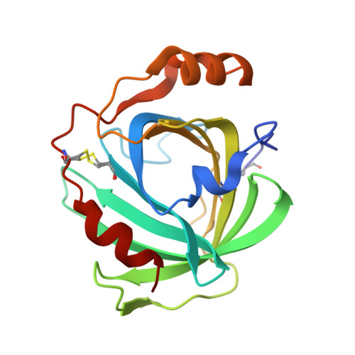
Guanidine-Ferroheme Coordination in the Mutant Protein Nitrophorin 4(L130R).

He, C.Fuchs, M.R.Ogata, H.Knipp, M.

(2012) Angew Chem Int Ed Engl 51: 4470-4473


Macromolecules
Find similar proteins by:  (by identity cutoff)  |  3D Structure
Entity ID: 1
MoleculeChains Sequence LengthOrganismDetailsImage
Nitrophorin-4184Rhodnius prolixusMutation(s): 1 
UniProt
Find proteins for Q94734 (Rhodnius prolixus)
Explore Q94734 
Go to UniProtKB:  Q94734
Entity Groups  
Sequence Clusters30% Identity50% Identity70% Identity90% Identity95% Identity100% Identity
UniProt GroupQ94734
Sequence Annotations
Expand
  • Reference Sequence
Small Molecules
Ligands 1 Unique
IDChains Name / Formula / InChI Key2D Diagram3D Interactions
HEM
Query on HEM

Download Ideal Coordinates CCD File 
B [auth A]PROTOPORPHYRIN IX CONTAINING FE
C34 H32 Fe N4 O4
KABFMIBPWCXCRK-RGGAHWMASA-L
Experimental Data & Validation

Experimental Data

  • Method: X-RAY DIFFRACTION
  • Resolution: 1.30 Å
  • R-Value Free: 0.172 
  • R-Value Work: 0.145 
  • R-Value Observed: 0.147 
  • Space Group: C 1 2 1
Unit Cell:
Length ( Å )Angle ( ˚ )
a = 69.89α = 90
b = 43.061β = 94.36
c = 52.585γ = 90
Software Package:
Software NamePurpose
XDSdata scaling
MOLREPphasing
PHENIXrefinement
XDSdata reduction
XSCALEdata scaling

Structure Validation

View Full Validation Report


Ligand Structure Quality Assessment 


Entry History 

Deposition Data

Revision History  (Full details and data files)

  • Version 1.0: 2012-05-30
    Type: Initial release