3TDK

Crystal Structure of Human UDP-Glucose Dehydrogenase


Experimental Data Snapshot

  • Method: X-RAY DIFFRACTION
  • Resolution: 2.80 Å
  • R-Value Free: 0.261 
  • R-Value Work: 0.230 
  • R-Value Observed: 0.231 

wwPDB Validation   3D Report Full Report


Ligand Structure Quality Assessment 


This is version 1.1 of the entry. See complete history


Literature

Structural Basis of Cooperativity in Human UDP-Glucose Dehydrogenase.

Rajakannan, V.Lee, H.S.Chong, S.H.Ryu, H.B.Bae, J.Y.Whang, E.Y.Huh, J.W.Cho, S.W.Kang, L.W.Choe, H.Robinson, R.C.

(2011) PLoS One 6: e25226-e25226

  • DOI: https://doi.org/10.1371/journal.pone.0025226
  • Primary Citation of Related Structures:  
    3TDK

  • PubMed Abstract: 

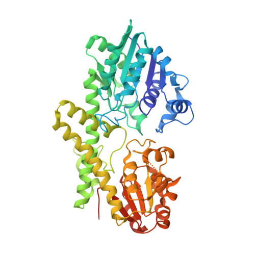
    UDP-glucose dehydrogenase (UGDH) is the sole enzyme that catalyzes the conversion of UDP-glucose to UDP-glucuronic acid. The product is used in xenobiotic glucuronidation in hepatocytes and in the production of proteoglycans that are involved in promoting normal cellular growth and migration. Overproduction of proteoglycans has been implicated in the progression of certain epithelial cancers, while inhibition of UGDH diminished tumor angiogenesis in vivo. A better understanding of the conformational changes occurring during the UGDH reaction cycle will pave the way for inhibitor design and potential cancer therapeutics. Previously, the substrate-bound of UGDH was determined to be a symmetrical hexamer and this regular symmetry is disrupted on binding the inhibitor, UDP-α-D-xylose. Here, we have solved an alternate crystal structure of human UGDH (hUGDH) in complex with UDP-glucose at 2.8 Å resolution. Surprisingly, the quaternary structure of this substrate-bound protein complex consists of the open homohexamer that was previously observed for inhibitor-bound hUGDH, indicating that this conformation is relevant for deciphering elements of the normal reaction cycle. In all subunits of the present open structure, Thr131 has translocated into the active site occupying the volume vacated by the absent active water and partially disordered NAD+ molecule. This conformation suggests a mechanism by which the enzyme may exchange NADH for NAD+ and repolarize the catalytic water bound to Asp280 while protecting the reaction intermediates. The structure also indicates how the subunits may communicate with each other through two reaction state sensors in this highly cooperative enzyme.


  • Organizational Affiliation

    Institute of Molecular and Cell Biology, Agency for Science, Technology and Research, Singapore, Singapore.


Macromolecules
Find similar proteins by:  (by identity cutoff)  |  3D Structure
Entity ID: 1
MoleculeChains Sequence LengthOrganismDetailsImage
UDP-glucose 6-dehydrogenase487Homo sapiensMutation(s): 0 
Gene Names: UGDH
EC: 1.1.1.22
UniProt & NIH Common Fund Data Resources
Find proteins for O60701 (Homo sapiens)
Explore O60701 
Go to UniProtKB:  O60701
PHAROS:  O60701
GTEx:  ENSG00000109814 
Entity Groups  
Sequence Clusters30% Identity50% Identity70% Identity90% Identity95% Identity100% Identity
UniProt GroupO60701
Sequence Annotations
Expand
  • Reference Sequence
Small Molecules
Ligands 2 Unique
IDChains Name / Formula / InChI Key2D Diagram3D Interactions
NAD
Query on NAD

Download Ideal Coordinates CCD File 
AA [auth E]
CA [auth D]
EA [auth C]
GA [auth J]
IA [auth I]
AA [auth E],
CA [auth D],
EA [auth C],
GA [auth J],
IA [auth I],
M [auth A],
O [auth B],
Q [auth G],
S [auth H],
U [auth L],
W [auth K],
Z [auth F]
NICOTINAMIDE-ADENINE-DINUCLEOTIDE
C21 H27 N7 O14 P2
BAWFJGJZGIEFAR-NNYOXOHSSA-N
UPG
Query on UPG

Download Ideal Coordinates CCD File 
BA [auth E]
DA [auth D]
FA [auth C]
HA [auth J]
JA [auth I]
BA [auth E],
DA [auth D],
FA [auth C],
HA [auth J],
JA [auth I],
N [auth A],
P [auth B],
R [auth G],
T [auth H],
V [auth L],
X [auth K],
Y [auth F]
URIDINE-5'-DIPHOSPHATE-GLUCOSE
C15 H24 N2 O17 P2
HSCJRCZFDFQWRP-JZMIEXBBSA-N
Experimental Data & Validation

Experimental Data

  • Method: X-RAY DIFFRACTION
  • Resolution: 2.80 Å
  • R-Value Free: 0.261 
  • R-Value Work: 0.230 
  • R-Value Observed: 0.231 
  • Space Group: P 21 21 21
Unit Cell:
Length ( Å )Angle ( ˚ )
a = 173.132α = 90
b = 191.177β = 90
c = 225.807γ = 90
Software Package:
Software NamePurpose
MOSFLMdata reduction
SCALAdata scaling
PHASERphasing
REFMACrefinement
PDB_EXTRACTdata extraction
ADSCdata collection

Structure Validation

View Full Validation Report


Ligand Structure Quality Assessment 


Entry History 

Deposition Data

Revision History  (Full details and data files)

  • Version 1.0: 2011-10-26
    Type: Initial release
  • Version 1.1: 2024-03-20
    Changes: Data collection, Database references, Derived calculations