3TDG

Structural and functional characterization of Helicobacter pylori DsbG


Experimental Data Snapshot

  • Method: X-RAY DIFFRACTION
  • Resolution: 2.10 Å
  • R-Value Free: 0.245 
  • R-Value Work: 0.220 
  • R-Value Observed: 0.222 

wwPDB Validation   3D Report Full Report


This is version 1.2 of the entry. See complete history


Literature

Structural and functional characterization of Helicobacter pylori DsbG

Yoon, J.Y.Kim, J.Lee, S.J.Kim, H.S.Im, H.N.Yoon, H.Kim, K.H.Kim, S.Han, B.W.Suh, S.W.

(2011) FEBS Lett 585: 3862-3867

  • DOI: https://doi.org/10.1016/j.febslet.2011.10.042
  • Primary Citation of Related Structures:  
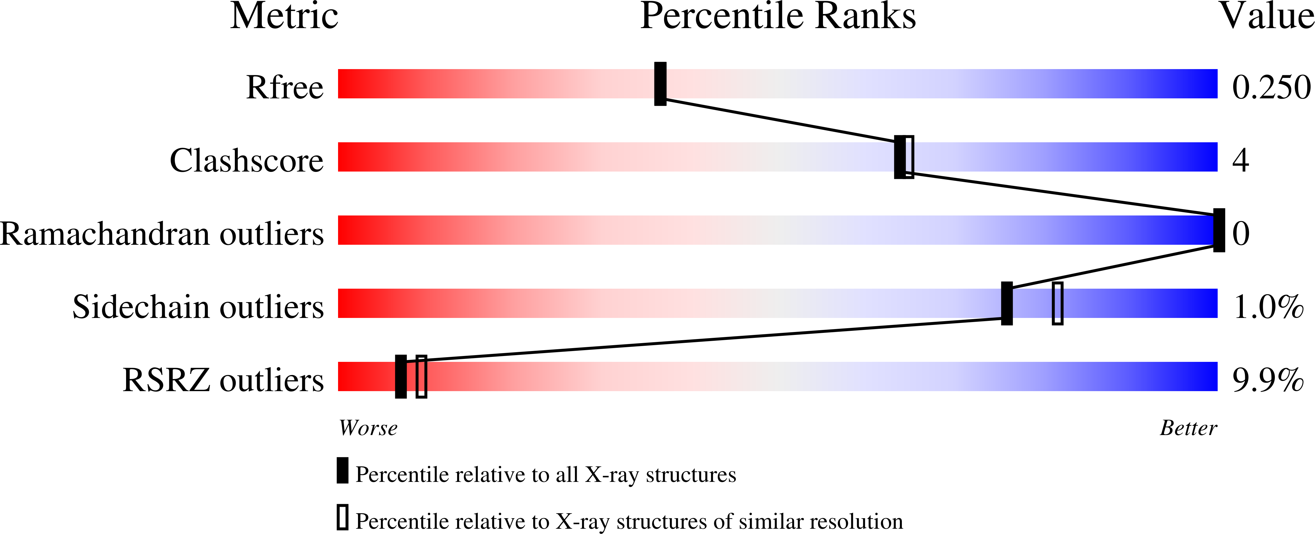
    3TDG

  • PubMed Abstract: 

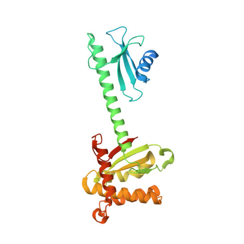
    Dsb proteins play important roles in bacterial pathogenicity. To better understand the role of Dsb proteins in Helicobacter pylori, we have structurally and functionally characterized H. pylori DsbG (HP0231). The monomer consists of two domains connected by a helical linker. Two monomers associate to form a V-shaped dimer. The monomeric and dimeric structures of H. pylori DsbG show significant differences compared to Escherichia coli DsbG. Two polyethylene glycol molecules are bound in the cleft of the V-shaped dimer, suggesting a possible role as a chaperone. Furthermore, we show that H. pylori DsbG functions as a reductase against HP0518, a putative L,D-transpeptidase with a catalytic cysteine residue.


  • Organizational Affiliation

    Department of Chemistry, College of Natural Sciences, Seoul National University, Seoul, Republic of Korea.


Macromolecules
Find similar proteins by:  (by identity cutoff)  |  3D Structure
Entity ID: 1
MoleculeChains Sequence LengthOrganismDetailsImage
Putative uncharacterized protein273Helicobacter pyloriMutation(s): 0 
Gene Names: HP_0231
UniProt
Find proteins for O25017 (Helicobacter pylori (strain ATCC 700392 / 26695))
Explore O25017 
Go to UniProtKB:  O25017
Entity Groups  
Sequence Clusters30% Identity50% Identity70% Identity90% Identity95% Identity100% Identity
UniProt GroupO25017
Sequence Annotations
Expand
  • Reference Sequence
Experimental Data & Validation

Experimental Data

  • Method: X-RAY DIFFRACTION
  • Resolution: 2.10 Å
  • R-Value Free: 0.245 
  • R-Value Work: 0.220 
  • R-Value Observed: 0.222 
  • Space Group: P 32 2 1
Unit Cell:
Length ( Å )Angle ( ˚ )
a = 86.767α = 90
b = 86.767β = 90
c = 85.508γ = 120
Software Package:
Software NamePurpose
HKL-2000data collection
SOLVEphasing
REFMACrefinement
HKL-2000data reduction
HKL-2000data scaling

Structure Validation

View Full Validation Report


Entry History 

Deposition Data

Revision History  (Full details and data files)

  • Version 1.0: 2011-11-09
    Type: Initial release
  • Version 1.1: 2013-07-03
    Changes: Database references
  • Version 1.2: 2024-03-20
    Changes: Data collection, Database references, Derived calculations