3TBC

Small laccase from Streptomyces viridosporus T7A; alternate crystal form complexed with acetovanillone.


Experimental Data Snapshot

  • Method: X-RAY DIFFRACTION
  • Resolution: 2.70 Å
  • R-Value Free: 0.210 
  • R-Value Work: 0.157 
  • R-Value Observed: 0.160 

wwPDB Validation   3D Report Full Report


Ligand Structure Quality Assessment 


This is version 1.4 of the entry. See complete history


Literature

Roles of small laccases from Streptomyces in lignin degradation.

Majumdar, S.Lukk, T.Solbiati, J.O.Bauer, S.Nair, S.K.Cronan, J.E.Gerlt, J.A.

(2014) Biochemistry 53: 4047-4058

  • DOI: https://doi.org/10.1021/bi500285t
  • Primary Citation of Related Structures:  
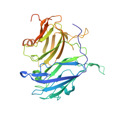
    3T9W, 3TA4, 3TAS, 3TBB, 3TBC

  • PubMed Abstract: 

    Laccases (EC 1.10.3.2) are multicopper oxidases that can oxidize a range of substrates, including phenols, aromatic amines, and nonphenolic substrates. To investigate the involvement of the small Streptomyces laccases in lignin degradation, we generated acid-precipitable polymeric lignin obtained in the presence of wild-type Streptomyces coelicolor A3(2) (SCWT) and its laccase-less mutant (SCΔLAC) in the presence of Miscanthus x giganteus lignocellulose. The results showed that strain SCΔLAC was inefficient in degrading lignin compared to strain SCWT, thereby supporting the importance of laccase for lignin degradation by S. coelicolor A3(2). We also studied the lignin degradation activity of laccases from S. coelicolor A3(2), Streptomyces lividans TK24, Streptomyces viridosporus T7A, and Amycolatopsis sp. 75iv2 using both lignin model compounds and ethanosolv lignin. All four laccases degraded a phenolic model compound (LM-OH) but were able to oxidize a nonphenolic model compound only in the presence of redox mediators. Their activities are highest at pH 8.0 with a low krel/Kapp for LM-OH, suggesting that the enzymes’ natural substrates must be different in shape or chemical nature. Crystal structures of the laccases from S. viridosporus T7A (SVLAC) and Amycolatopsis sp. 75iv2 were determined both with and without bound substrate. This is the first report of a crystal structure for any laccase bound to a nonphenolic β-O-4 lignin model compound. An additional zinc metal binding site in SVLAC was also identified. The ability to oxidize and/or rearrange ethanosolv lignin provides further evidence of the utility of laccase activity for lignin degradation and/or modification.


Macromolecules
Find similar proteins by:  (by identity cutoff)  |  3D Structure
Entity ID: 1
MoleculeChains Sequence LengthOrganismDetailsImage
small laccase, two-domain laccase, multi-copper oxidase
A, B, C
313Streptomyces viridosporusMutation(s): 0 
EC: 1.10.3.2
UniProt
Find proteins for J9PBR2 (Streptomyces viridosporus)
Explore J9PBR2 
Go to UniProtKB:  J9PBR2
Entity Groups  
Sequence Clusters30% Identity50% Identity70% Identity90% Identity95% Identity100% Identity
UniProt GroupJ9PBR2
Sequence Annotations
Expand
  • Reference Sequence
Small Molecules
Ligands 5 Unique
IDChains Name / Formula / InChI Key2D Diagram3D Interactions
I75
Query on I75

Download Ideal Coordinates CCD File 
W [auth C]1-(4-hydroxy-3-methoxyphenyl)ethanone
C9 H10 O3
DFYRUELUNQRZTB-UHFFFAOYSA-N
TAM
Query on TAM

Download Ideal Coordinates CCD File 
X [auth C]TRIS(HYDROXYETHYL)AMINOMETHANE
C7 H17 N O3
GKODZWOPPOTFGA-UHFFFAOYSA-N
PO4
Query on PO4

Download Ideal Coordinates CCD File 
H [auth A]
I [auth A]
O [auth B]
P [auth B]
Q [auth B]
H [auth A],
I [auth A],
O [auth B],
P [auth B],
Q [auth B],
Y [auth C],
Z [auth C]
PHOSPHATE ION
O4 P
NBIIXXVUZAFLBC-UHFFFAOYSA-K
CU
Query on CU

Download Ideal Coordinates CCD File 
D [auth A]
E [auth A]
F [auth A]
J [auth B]
K [auth B]
D [auth A],
E [auth A],
F [auth A],
J [auth B],
K [auth B],
L [auth B],
M [auth B],
N [auth B],
R [auth C],
S [auth C],
T [auth C],
U [auth C]
COPPER (II) ION
Cu
JPVYNHNXODAKFH-UHFFFAOYSA-N
OXY
Query on OXY

Download Ideal Coordinates CCD File 
G [auth A],
V [auth C]
OXYGEN MOLECULE
O2
MYMOFIZGZYHOMD-UHFFFAOYSA-N
Experimental Data & Validation

Experimental Data

  • Method: X-RAY DIFFRACTION
  • Resolution: 2.70 Å
  • R-Value Free: 0.210 
  • R-Value Work: 0.157 
  • R-Value Observed: 0.160 
  • Space Group: P 31 2 1
Unit Cell:
Length ( Å )Angle ( ˚ )
a = 127.02α = 90
b = 127.02β = 90
c = 155.54γ = 120
Software Package:
Software NamePurpose
XSCALEdata scaling
PHENIXrefinement
PDB_EXTRACTdata extraction
HKL-2000data collection
XDSdata reduction
PHASERphasing

Structure Validation

View Full Validation Report


Ligand Structure Quality Assessment 


Entry History 

Deposition Data

Revision History  (Full details and data files)

  • Version 1.0: 2012-08-29
    Type: Initial release
  • Version 1.1: 2014-06-11
    Changes: Database references
  • Version 1.2: 2014-10-01
    Changes: Database references
  • Version 1.3: 2017-10-25
    Changes: Author supporting evidence
  • Version 1.4: 2024-02-28
    Changes: Data collection, Database references, Derived calculations