


Crystal Structure of diiron adenine deaminase
Bagaria, A., Kumaran, D., Burley, S.K., Swaminathan, S.To be published.
Experimental Data Snapshot
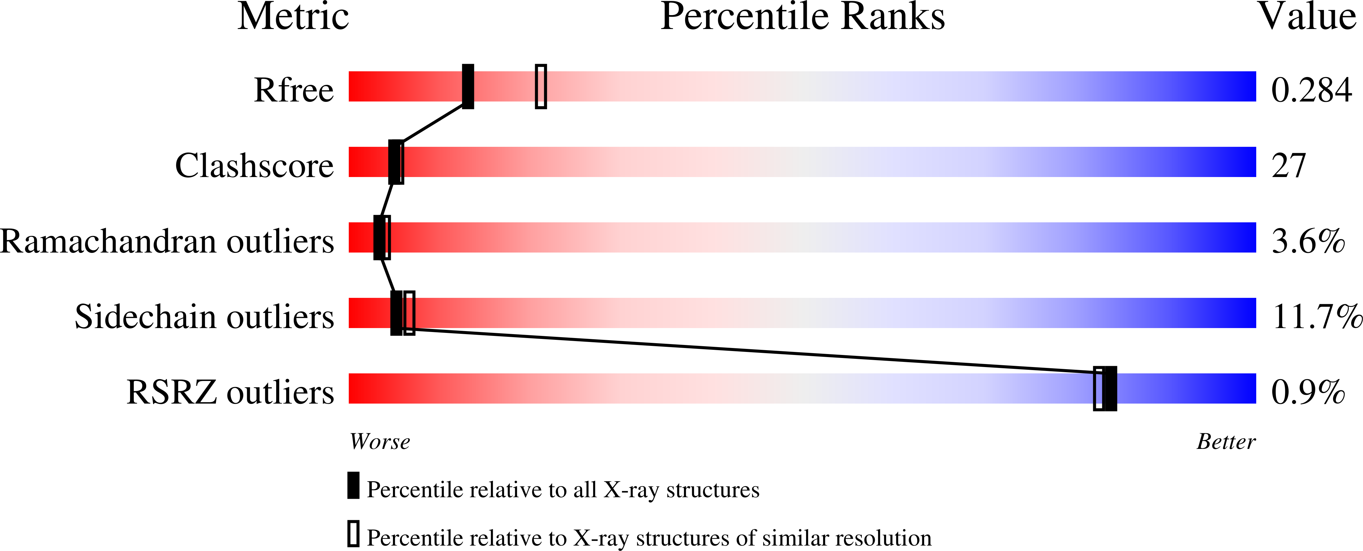
wwPDB Validation 3D Report Full Report
Entity ID: 1 | |||||
|---|---|---|---|---|---|
| Molecule | Chains | Sequence Length | Organism | Details | Image |
| Adenine deaminase 2 | 608 | Agrobacterium fabrum str. C58 | Mutation(s): 0 Gene Names: ade2, adeC, AGR_L_883, Atu4426 EC: 3.5.4.2 | 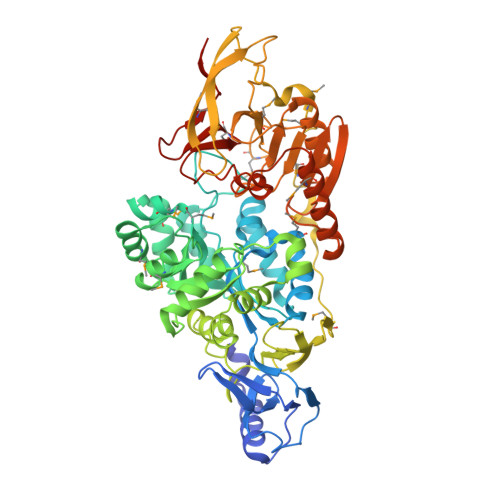 | |
UniProt | |||||
Find proteins for Q7CUX4 (Agrobacterium fabrum (strain C58 / ATCC 33970)) Explore Q7CUX4 Go to UniProtKB: Q7CUX4 | |||||
Entity Groups | |||||
| Sequence Clusters | 30% Identity50% Identity70% Identity90% Identity95% Identity100% Identity | ||||
| UniProt Group | Q7CUX4 | ||||
Sequence AnnotationsExpand | |||||
| |||||
| Ligands 1 Unique | |||||
|---|---|---|---|---|---|
| ID | Chains | Name / Formula / InChI Key | 2D Diagram | 3D Interactions | |
| FE Query on FE | C [auth A] D [auth A] E [auth A] F [auth B] G [auth B] | FE (III) ION Fe VTLYFUHAOXGGBS-UHFFFAOYSA-N |  | ||
| Modified Residues 1 Unique | |||||
|---|---|---|---|---|---|
| ID | Chains | Type | Formula | 2D Diagram | Parent |
| MSE Query on MSE | A, B | L-PEPTIDE LINKING | C5 H11 N O2 Se |  | MET |
| Length ( Å ) | Angle ( ˚ ) |
|---|---|
| a = 61.662 | α = 90 |
| b = 131.845 | β = 97.04 |
| c = 69.628 | γ = 90 |
| Software Name | Purpose |
|---|---|
| CBASS | data collection |
| REFMAC | refinement |
| HKL-2000 | data reduction |
| HKL-2000 | data scaling |
| REFMAC | phasing |
RCSB PDB (citation) is hosted by
RCSB PDB is a member of the

