3T50

X-ray structure of the LOV domain from the LOV-HK sensory protein from Brucella abortus (dark state).


Experimental Data Snapshot

  • Method: X-RAY DIFFRACTION
  • Resolution: 1.64 Å
  • R-Value Free: 0.222 
  • R-Value Work: 0.193 
  • R-Value Observed: 0.195 

wwPDB Validation   3D Report Full Report


Ligand Structure Quality Assessment 


This is version 1.1 of the entry. See complete history


Literature

The Beta-Scaffold of the LOV Domain of the Brucella Light-Activated Histidine Kinase Is a Key Element for Signal Transduction

Rinaldi, J.Gallo, M.Klinke, S.Paris, G.Bonomi, H.R.Bogomolni, R.A.Cicero, D.O.Goldbaum, F.A.

(2012) J Mol Biol 420: 112-127

  • DOI: https://doi.org/10.1016/j.jmb.2012.04.006
  • Primary Citation of Related Structures:  
    3T50

  • PubMed Abstract: 

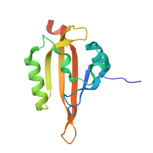
    Light-oxygen-voltage (LOV) domains are blue-light-activated signaling modules present in a wide range of sensory proteins. Among them, the histidine kinases are the largest group in prokaryotes (LOV-HK). Light modulates the virulence of the pathogenic bacteria Brucella abortus through LOV-HK. One of the striking characteristic of Brucella LOV-HK is the fact that the protein remains activated upon light sensing, without recovering the basal state in the darkness. In contrast, the light state of the isolated LOV domain slowly returns to the dark state. To gain insight into the light activation mechanism, we have characterized by X-ray crystallography and solution NMR spectroscopy the structure of the LOV domain of LOV-HK in the dark state and explored its light-induced conformational changes. The LOV domain adopts the α/β PAS (PER-ARNT-SIM) domain fold and binds the FMN cofactor within a conserved pocket. The domain dimerizes through the hydrophobic β-scaffold in an antiparallel way. Our results point to the β-scaffold as a key element in the light activation, validating a conserved structural basis for light-to-signal propagation in LOV proteins.


  • Organizational Affiliation

    Fundación Instituto Leloir, IIBBA-CONICET, Ciudad Autónoma de Buenos Aires, Buenos Aires, Argentina.


Macromolecules
Find similar proteins by:  (by identity cutoff)  |  3D Structure
Entity ID: 1
MoleculeChains Sequence LengthOrganismDetailsImage
Blue-light-activated histidine kinase
A, B
128Brucella melitensisMutation(s): 0 
Gene Names: BAB2_0652BMEII0679
EC: 2.7.13.3
UniProt
Find proteins for Q8YC53 (Brucella melitensis biotype 1 (strain 16M / ATCC 23456 / NCTC 10094))
Explore Q8YC53 
Go to UniProtKB:  Q8YC53
Entity Groups  
Sequence Clusters30% Identity50% Identity70% Identity90% Identity95% Identity100% Identity
UniProt GroupQ8YC53
Sequence Annotations
Expand
  • Reference Sequence
Small Molecules
Experimental Data & Validation

Experimental Data

  • Method: X-RAY DIFFRACTION
  • Resolution: 1.64 Å
  • R-Value Free: 0.222 
  • R-Value Work: 0.193 
  • R-Value Observed: 0.195 
  • Space Group: P 21 21 21
Unit Cell:
Length ( Å )Angle ( ˚ )
a = 37.9α = 90
b = 61.6β = 90
c = 98.6γ = 90
Software Package:
Software NamePurpose
MAR345dtbdata collection
PHASERphasing
REFMACrefinement
XDSdata reduction
XDSdata scaling

Structure Validation

View Full Validation Report


Ligand Structure Quality Assessment 


Entry History 

Deposition Data

Revision History  (Full details and data files)

  • Version 1.0: 2012-06-13
    Type: Initial release
  • Version 1.1: 2023-09-13
    Changes: Data collection, Database references, Derived calculations, Refinement description