3SVL

Structural basis of the improvement of ChrR - a multi-purpose enzyme


Experimental Data Snapshot

  • Method: X-RAY DIFFRACTION
  • Resolution: 2.20 Å
  • R-Value Free: 0.270 
  • R-Value Work: 0.237 
  • R-Value Observed: 0.257 

wwPDB Validation   3D Report Full Report


Ligand Structure Quality Assessment 


This is version 1.1 of the entry. See complete history


Literature

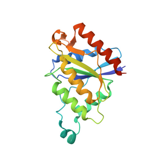
Crystal Structure of ChrR-A Quinone Reductase with the Capacity to Reduce Chromate.

Eswaramoorthy, S.Poulain, S.Hienerwadel, R.Bremond, N.Sylvester, M.D.Zhang, Y.B.Berthomieu, C.Van Der Lelie, D.Matin, A.

(2012) PLoS One 7: e36017-e36017

  • DOI: https://doi.org/10.1371/journal.pone.0036017
  • Primary Citation of Related Structures:  
    3SVL

  • PubMed Abstract: 

    The Escherichia coli ChrR enzyme is an obligatory two-electron quinone reductase that has many applications, such as in chromate bioremediation. Its crystal structure, solved at 2.2 Å resolution, shows that it belongs to the flavodoxin superfamily in which flavin mononucleotide (FMN) is firmly anchored to the protein. ChrR crystallized as a tetramer, and size exclusion chromatography showed that this is the oligomeric form that catalyzes chromate reduction. Within the tetramer, the dimers interact by a pair of two hydrogen bond networks, each involving Tyr128 and Glu146 of one dimer and Arg125 and Tyr85 of the other; the latter extends to one of the redox FMN cofactors. Changes in each of these amino acids enhanced chromate reductase activity of the enzyme, showing that this network is centrally involved in chromate reduction.


  • Organizational Affiliation

    Department of Biology, Brookhaven National Laboratory, Upton, New York, USA.


Macromolecules
Find similar proteins by:  (by identity cutoff)  |  3D Structure
Entity ID: 1
MoleculeChains Sequence LengthOrganismDetailsImage
protein yieF
A, B
193Escherichia coli K-12Mutation(s): 0 
Gene Names: yieFb3713JW3691
UniProt
Find proteins for P0AGE6 (Escherichia coli (strain K12))
Explore P0AGE6 
Go to UniProtKB:  P0AGE6
Entity Groups  
Sequence Clusters30% Identity50% Identity70% Identity90% Identity95% Identity100% Identity
UniProt GroupP0AGE6
Sequence Annotations
Expand
  • Reference Sequence
Experimental Data & Validation

Experimental Data

  • Method: X-RAY DIFFRACTION
  • Resolution: 2.20 Å
  • R-Value Free: 0.270 
  • R-Value Work: 0.237 
  • R-Value Observed: 0.257 
  • Space Group: P 63 2 2
Unit Cell:
Length ( Å )Angle ( ˚ )
a = 107.255α = 90
b = 107.255β = 90
c = 128.251γ = 120
Software Package:
Software NamePurpose
CBASSdata collection
MOLREPphasing
CNSrefinement
HKL-2000data reduction
HKL-2000data scaling

Structure Validation

View Full Validation Report


Ligand Structure Quality Assessment 


Entry History 

Deposition Data

Revision History  (Full details and data files)

  • Version 1.0: 2012-05-30
    Type: Initial release
  • Version 1.1: 2023-09-13
    Changes: Data collection, Database references, Derived calculations, Refinement description