3SM8

Crystal Structure of Pseudomonas aeruginosa D-Arginine Dehydrogenase in Complex with an (N5) Flavin Adduct


Experimental Data Snapshot

  • Method: X-RAY DIFFRACTION
  • Resolution: 1.07 Å
  • R-Value Free: 0.165 
  • R-Value Work: 0.145 
  • R-Value Observed: 0.145 

wwPDB Validation   3D Report Full Report


Ligand Structure Quality Assessment 


This is version 1.3 of the entry. See complete history


Literature

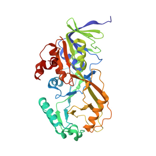
Atomic-resolution structure of an N5 flavin adduct in D-arginine dehydrogenase.

Fu, G.Yuan, H.Wang, S.Gadda, G.Weber, I.T.

(2011) Biochemistry 50: 6292-6294

  • DOI: https://doi.org/10.1021/bi200831a
  • Primary Citation of Related Structures:  
    3SM8

  • PubMed Abstract: 

    D-Arginine dehydrogenase (DADH) catalyzes the flavin-dependent oxidative deamination of D-arginine and other D-amino acids to the corresponding imino acids. The 1.07 Å atomic-resolution structure of DADH crystallized with D-leucine unexpectedly revealed a covalent N(5) flavin adduct, instead of the expected iminoleucine product in the active site. This acyl adduct has been successfully reproduced by photoreduction of DADH in the presence of 4-methyl-2-oxopentanoic acid (ketoleucine). The iminoleucine may be released readily because of weak interactions in the binding site, in contrast to iminoarginine, converted to ketoleucine, which reacts with activated FAD to form the covalently linked acyl adduct.


  • Organizational Affiliation

    Department of Biology, Georgia State University, Atlanta, Georgia 30303, USA.


Macromolecules
Find similar proteins by:  (by identity cutoff)  |  3D Structure
Entity ID: 1
MoleculeChains Sequence LengthOrganismDetailsImage
FAD-dependent catabolic D-arginine dehydrogenase, DauA381Pseudomonas aeruginosaMutation(s): 0 
Gene Names: dauAPA3863PAO1
EC: 1.4.1
UniProt
Find proteins for Q9HXE3 (Pseudomonas aeruginosa (strain ATCC 15692 / DSM 22644 / CIP 104116 / JCM 14847 / LMG 12228 / 1C / PRS 101 / PAO1))
Explore Q9HXE3 
Go to UniProtKB:  Q9HXE3
Entity Groups  
Sequence Clusters30% Identity50% Identity70% Identity90% Identity95% Identity100% Identity
UniProt GroupQ9HXE3
Sequence Annotations
Expand
  • Reference Sequence
Small Molecules
Ligands 2 Unique
IDChains Name / Formula / InChI Key2D Diagram3D Interactions
FNK
Query on FNK

Download Ideal Coordinates CCD File 
E [auth A][(2R,3S,4R,5R)-5-(6-amino-9H-purin-9-yl)-3,4-dihydroxytetrahydrofuran-2-yl]methyl (2R,3S,4S)-5-[7,8-dimethyl-5-(3-methylbutanoyl)-2,4-dioxo-1,3,4,5-tetrahydrobenzo[g]pteridin-10(2H)-yl]-2,3,4-trihydroxypentyl dihydrogen diphosphate
C32 H43 N9 O16 P2
CXENQOVRLDMZEP-KQGUAMSLSA-N
GOL
Query on GOL

Download Ideal Coordinates CCD File 
B [auth A],
C [auth A],
D [auth A]
GLYCEROL
C3 H8 O3
PEDCQBHIVMGVHV-UHFFFAOYSA-N
Experimental Data & Validation

Experimental Data

  • Method: X-RAY DIFFRACTION
  • Resolution: 1.07 Å
  • R-Value Free: 0.165 
  • R-Value Work: 0.145 
  • R-Value Observed: 0.145 
  • Space Group: P 21 21 21
Unit Cell:
Length ( Å )Angle ( ˚ )
a = 62.072α = 90
b = 77.933β = 90
c = 89.31γ = 90
Software Package:
Software NamePurpose
HKL-2000data collection
PHASERphasing
SHELXL-97refinement
HKL-2000data reduction
HKL-2000data scaling

Structure Validation

View Full Validation Report


Ligand Structure Quality Assessment 


Entry History 

Deposition Data

Revision History  (Full details and data files)

  • Version 1.0: 2011-07-20
    Type: Initial release
  • Version 1.1: 2012-01-11
    Changes: Database references
  • Version 1.2: 2024-02-28
    Changes: Data collection, Database references, Derived calculations
  • Version 1.3: 2024-04-03
    Changes: Refinement description