3SHM

Structure-function Analysis of Receptor Binding in Adeno-Associated Virus Serotype 6 (AAV-6)


Experimental Data Snapshot

  • Method: X-RAY DIFFRACTION
  • Resolution: 3.02 Å
  • R-Value Free: 0.286 
  • R-Value Work: 0.273 
  • R-Value Observed: 0.273 

wwPDB Validation   3D Report Full Report


This is version 1.4 of the entry. See complete history


Literature

Structure-function analysis of receptor-binding in adeno-associated virus serotype 6 (AAV-6).

Xie, Q.Lerch, T.F.Meyer, N.L.Chapman, M.S.

(2011) Virology 420: 10-19

  • DOI: https://doi.org/10.1016/j.virol.2011.08.011
  • Primary Citation of Related Structures:  
    3SHM, 4V86

  • PubMed Abstract: 

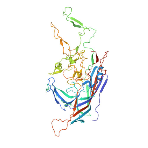
    Crystal structures of the AAV-6 capsid at 3Å reveal a subunit fold homologous to other parvoviruses with greatest differences in two external loops. The electrostatic potential suggests that receptor-attachment is mediated by four residues: Arg(576), Lys(493), Lys(459) and Lys(531), defining a positively charged region curving up from the valley between adjacent spikes. It overlaps only partially with the receptor-binding site of AAV-2, and the residues endowing the electrostatic character are not homologous. Mutational substitution of each residue decreases heparin affinity, particularly Lys(531) and Lys(459). Neither is conserved among heparin-binding serotypes, indicating that diverse modes of receptor attachment have been selected in different serotypes. Surface topology and charge are also distinct at the shoulder of the spike, where linear epitopes for AAV-2's neutralizing monoclonal antibody A20 come together. Evolutionarily, selection of changed side-chain charge may have offered a conservative means to evade immune neutralization while preserving other essential functionality.


  • Organizational Affiliation

    Department of Biochemistry & Molecular Biology, School of Medicine, Oregon Health & Science University, Portland, OR 97239-3098, USA.


Macromolecules
Find similar proteins by:  (by identity cutoff)  |  3D Structure
Entity ID: 1
MoleculeChains Sequence LengthOrganismDetailsImage
Capsid protein VP1
A, B, C, D, E
A, B, C, D, E, F, G, H, I, J, K, L, M, N, O, P, Q, R, S, T
516Adeno-associated virus - 6Mutation(s): 0 
UniProt
Find proteins for O56137 (Adeno-associated virus - 6)
Explore O56137 
Go to UniProtKB:  O56137
Entity Groups  
Sequence Clusters30% Identity50% Identity70% Identity90% Identity95% Identity100% Identity
UniProt GroupO56137
Sequence Annotations
Expand
  • Reference Sequence
Experimental Data & Validation

Experimental Data

  • Method: X-RAY DIFFRACTION
  • Resolution: 3.02 Å
  • R-Value Free: 0.286 
  • R-Value Work: 0.273 
  • R-Value Observed: 0.273 
  • Space Group: H 3
Unit Cell:
Length ( Å )Angle ( ˚ )
a = 258.357α = 90
b = 258.357β = 90
c = 612.966γ = 120
Software Package:
Software NamePurpose
SCALEPACKdata scaling
PHENIXrefinement
PDB_EXTRACTdata extraction
ADSCdata collection
DENZOdata reduction
GLRFphasing

Structure Validation

View Full Validation Report


Entry History 

Deposition Data

Revision History  (Full details and data files)

  • Version 1.0: 2011-10-05
    Type: Initial release
  • Version 1.1: 2011-10-19
    Changes: Database references
  • Version 1.2: 2017-11-08
    Changes: Refinement description
  • Version 1.3: 2024-02-28
    Changes: Data collection, Database references, Derived calculations
  • Version 1.4: 2024-04-03
    Changes: Refinement description