3SEO

Crystal structure of VopL C terminal domain


Experimental Data Snapshot

  • Method: X-RAY DIFFRACTION
  • Resolution: 2.31 Å
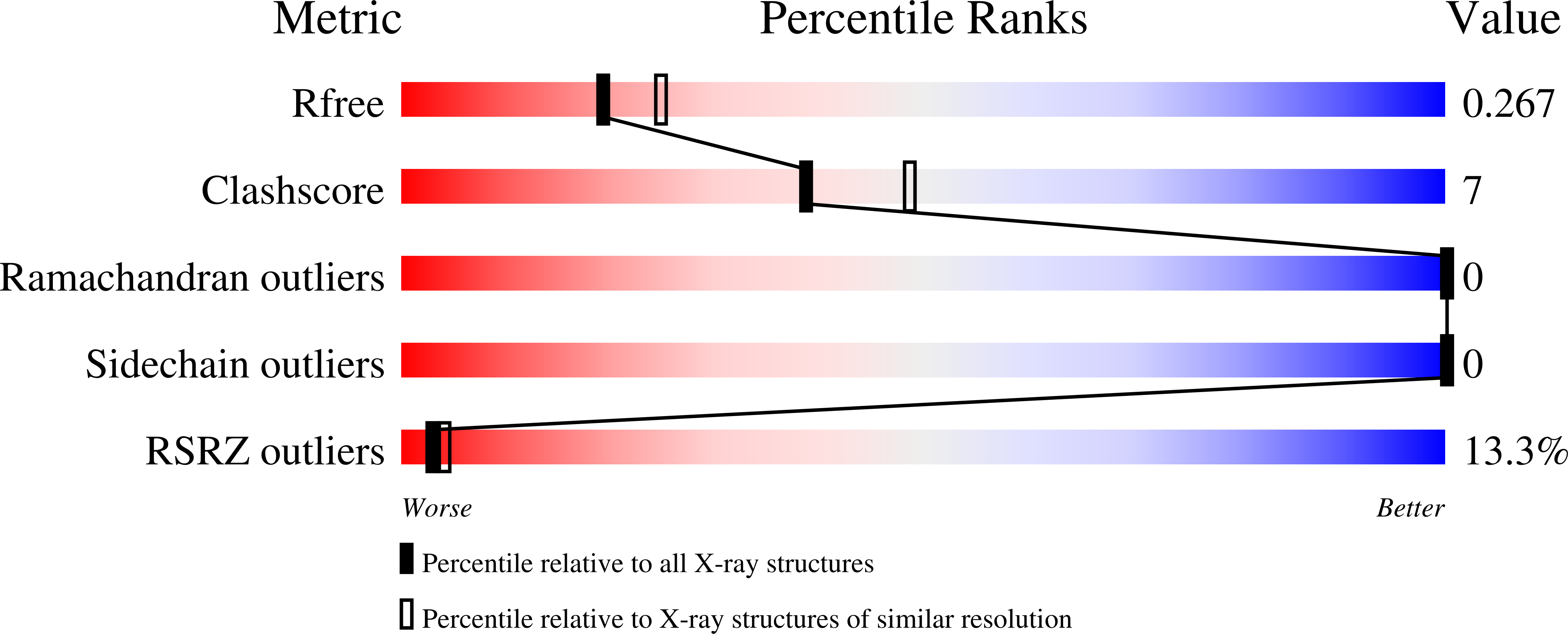
  • R-Value Free: 0.275 
  • R-Value Work: 0.221 
  • R-Value Observed: 0.225 

wwPDB Validation   3D Report Full Report


This is version 1.3 of the entry. See complete history


Literature

Mechanism of actin filament nucleation by the bacterial effector VopL.

Yu, B.Cheng, H.C.Brautigam, C.A.Tomchick, D.R.Rosen, M.K.

(2011) Nat Struct Mol Biol 18: 1068-1074

  • DOI: https://doi.org/10.1038/nsmb.2110
  • Primary Citation of Related Structures:  
    3SEO

  • PubMed Abstract: 

    Vibrio parahaemolyticus protein L (VopL) is an actin nucleation factor that induces stress fibers when injected into eukaryotic host cells. VopL contains three N-terminal Wiskott-Aldrich homology 2 (WH2) motifs and a unique VopL C-terminal domain (VCD). We describe crystallographic and biochemical analyses of filament nucleation by VopL. The WH2 element of VopL does not nucleate on its own and requires the VCD for activity. The VCD forms a U-shaped dimer in the crystal, stabilized by a terminal coiled coil. Dimerization of the WH2 motifs contributes strongly to nucleation activity, as do contacts of the VCD to actin. Our data lead to a model in which VopL stabilizes primarily lateral (short-pitch) contacts between actin monomers to create the base of a two-stranded filament. Stabilization of lateral contacts may be a common feature of actin filament nucleation by WH2-based factors.


  • Organizational Affiliation

    Department of Biochemistry, University of Texas Southwestern Medical Center, Dallas, Texas, USA.


Macromolecules
Find similar proteins by:  (by identity cutoff)  |  3D Structure
Entity ID: 1
MoleculeChains Sequence LengthOrganismDetailsImage
VopL C terminal domain protein
A, B
241Vibrio parahaemolyticusMutation(s): 0 
Gene Names: VopLVPA1370
UniProt
Find proteins for Q87GE5 (Vibrio parahaemolyticus serotype O3:K6 (strain RIMD 2210633))
Explore Q87GE5 
Go to UniProtKB:  Q87GE5
Entity Groups  
Sequence Clusters30% Identity50% Identity70% Identity90% Identity95% Identity100% Identity
UniProt GroupQ87GE5
Sequence Annotations
Expand
  • Reference Sequence
Experimental Data & Validation

Experimental Data

  • Method: X-RAY DIFFRACTION
  • Resolution: 2.31 Å
  • R-Value Free: 0.275 
  • R-Value Work: 0.221 
  • R-Value Observed: 0.225 
  • Space Group: P 21 21 21
Unit Cell:
Length ( Å )Angle ( ˚ )
a = 55.18α = 90
b = 89.851β = 90
c = 103.007γ = 90
Software Package:
Software NamePurpose
DENZOdata reduction
SCALEPACKdata scaling
PHENIXrefinement
PDB_EXTRACTdata extraction
HKL-3000data collection
HKL-3000data reduction
HKL-3000data scaling
SOLVEphasing

Structure Validation

View Full Validation Report


Entry History 

Deposition Data

Revision History  (Full details and data files)

  • Version 1.0: 2011-08-31
    Type: Initial release
  • Version 1.1: 2012-05-23
    Changes: Database references
  • Version 1.2: 2017-11-08
    Changes: Advisory, Refinement description
  • Version 1.3: 2024-02-28
    Changes: Advisory, Data collection, Database references, Derived calculations