3S9C

Russell's viper venom serine proteinase, RVV-V in complex with the fragment (residues 1533-1546) of human factor V


Experimental Data Snapshot

  • Method: X-RAY DIFFRACTION
  • Resolution: 1.80 Å
  • R-Value Free: 0.218 
  • R-Value Work: 0.183 
  • R-Value Observed: 0.183 

wwPDB Validation   3D Report Full Report


Ligand Structure Quality Assessment 


This is version 2.1 of the entry. See complete history


Literature

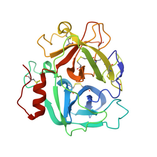
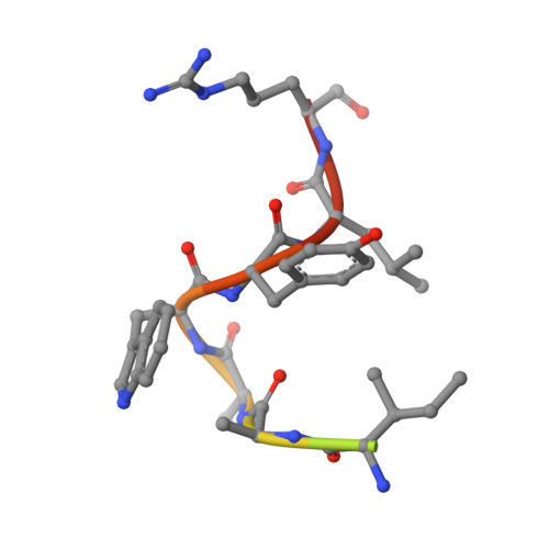
Structural basis of coagulation factor V recognition for cleavage by RVV-V

Nakayama, D.Ben Ammar, Y.Miyata, T.Takeda, S.

(2011) FEBS Lett 585: 3020-3025

  • DOI: https://doi.org/10.1016/j.febslet.2011.08.022
  • Primary Citation of Related Structures:  
    3S9A, 3S9B, 3S9C, 3SBK

  • PubMed Abstract: 

    Russell's viper venom factor V (FV) activator (RVV-V) is a thrombin-like proteinase that specifically cleaves the Arg1545-Ser1546 bond of FV. Here we present the crystal structure of RVV-V in complex with the FV14 peptide (residues 1533-1546 of human FV) determined at 1.8Å resolution. The structure reveals multiple interactions between RVV-V and the seven residues, Ile1539 (P(7))-Arg1545 (P(1)), of the cleaved substrate. Comparison with substrate-free structures reveals conformational changes of the RVV-V loops upon substrate binding, suggesting that the multiple interactions are mediated by an induced-fit mechanism. The results provide an explanation for the narrow specificity of RVV-V.


  • Organizational Affiliation

    Department of Cardiac Physiology, National Cerebral and Cardiovascular Center Research Institute, Suita, Osaka, Japan.


Macromolecules
Find similar proteins by:  (by identity cutoff)  |  3D Structure
Entity ID: 1
MoleculeChains Sequence LengthOrganismDetailsImage
Vipera russelli proteinase RVV-V gamma234Daboia siamensisMutation(s): 0 
EC: 3.4.21.95
UniProt
Find proteins for P18965 (Daboia siamensis)
Explore P18965 
Go to UniProtKB:  P18965
Entity Groups  
Sequence Clusters30% Identity50% Identity70% Identity90% Identity95% Identity100% Identity
UniProt GroupP18965
Sequence Annotations
Expand
  • Reference Sequence

Find similar proteins by:  Sequence   |   3D Structure  

Entity ID: 2
MoleculeChains Sequence LengthOrganismDetailsImage
Coagulation factor V14Homo sapiensMutation(s): 0 
UniProt & NIH Common Fund Data Resources
Find proteins for P12259 (Homo sapiens)
Explore P12259 
Go to UniProtKB:  P12259
PHAROS:  P12259
GTEx:  ENSG00000198734 
Entity Groups  
Sequence Clusters30% Identity50% Identity70% Identity90% Identity95% Identity100% Identity
UniProt GroupP12259
Sequence Annotations
Expand
  • Reference Sequence
Oligosaccharides

Help

Entity ID: 3
MoleculeChains Length2D Diagram Glycosylation3D Interactions
beta-D-mannopyranose-(1-4)-2-acetamido-2-deoxy-beta-D-glucopyranose-(1-4)-2-acetamido-2-deoxy-beta-D-glucopyranose
C
3N-Glycosylation
Glycosylation Resources
GlyTouCan:  G15407YE
GlyCosmos:  G15407YE
GlyGen:  G15407YE
Small Molecules
Ligands 4 Unique
IDChains Name / Formula / InChI Key2D Diagram3D Interactions
GLC
Query on GLC

Download Ideal Coordinates CCD File 
E [auth A]alpha-D-glucopyranose
C6 H12 O6
WQZGKKKJIJFFOK-DVKNGEFBSA-N
BGC
Query on BGC

Download Ideal Coordinates CCD File 
D [auth A]beta-D-glucopyranose
C6 H12 O6
WQZGKKKJIJFFOK-VFUOTHLCSA-N
ZN
Query on ZN

Download Ideal Coordinates CCD File 
J [auth A],
K [auth A],
L [auth A]
ZINC ION
Zn
PTFCDOFLOPIGGS-UHFFFAOYSA-N
ACT
Query on ACT

Download Ideal Coordinates CCD File 
F [auth A],
G [auth A],
H [auth A],
I [auth A]
ACETATE ION
C2 H3 O2
QTBSBXVTEAMEQO-UHFFFAOYSA-M
Experimental Data & Validation

Experimental Data

  • Method: X-RAY DIFFRACTION
  • Resolution: 1.80 Å
  • R-Value Free: 0.218 
  • R-Value Work: 0.183 
  • R-Value Observed: 0.183 
  • Space Group: P 61
Unit Cell:
Length ( Å )Angle ( ˚ )
a = 101.2α = 90
b = 101.2β = 90
c = 44.2γ = 120
Software Package:
Software NamePurpose
CNSrefinement
HKL-2000data collection
HKL-2000data reduction
HKL-2000data scaling
MOLREPphasing

Structure Validation

View Full Validation Report


Ligand Structure Quality Assessment 


Entry History 

Deposition Data

Revision History  (Full details and data files)

  • Version 1.0: 2011-09-07
    Type: Initial release
  • Version 1.1: 2013-07-03
    Changes: Database references
  • Version 2.0: 2020-07-29
    Type: Remediation
    Reason: Carbohydrate remediation
    Changes: Atomic model, Data collection, Database references, Derived calculations, Structure summary
  • Version 2.1: 2023-11-01
    Changes: Data collection, Database references, Refinement description, Structure summary