


Crystal structure of a Polysaccharide deacetylase family protein from Burkholderia pseudomallei
Seattle Structural Genomics Center for Infectious Disease (SSGCID), Gardberg, A., Edwards, T., Sankaran, B., Staker, B., Stewart, L.To be published.
Experimental Data Snapshot
wwPDB Validation 3D Report Full Report

Entity ID: 1 | |||||
|---|---|---|---|---|---|
| Molecule | Chains | Sequence Length | Organism | Details | Image |
| Polysaccharide deacetylase family protein | 321 | Burkholderia pseudomallei | Mutation(s): 0 Gene Names: BURPS1710b_2533 EC: 3.5.1 | 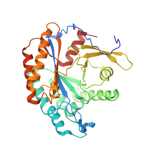 | |
UniProt | |||||
Find proteins for Q63T51 (Burkholderia pseudomallei (strain K96243)) Explore Q63T51 Go to UniProtKB: Q63T51 | |||||
Entity Groups | |||||
| Sequence Clusters | 30% Identity50% Identity70% Identity90% Identity95% Identity100% Identity | ||||
| UniProt Group | Q63T51 | ||||
Sequence AnnotationsExpand | |||||
| |||||
| Ligands 1 Unique | |||||
|---|---|---|---|---|---|
| ID | Chains | Name / Formula / InChI Key | 2D Diagram | 3D Interactions | |
| EDO Query on EDO | E [auth B], F [auth B] | 1,2-ETHANEDIOL C2 H6 O2 LYCAIKOWRPUZTN-UHFFFAOYSA-N |  | ||
| Length ( Å ) | Angle ( ˚ ) |
|---|---|
| a = 51.41 | α = 90 |
| b = 165.66 | β = 90 |
| c = 165.84 | γ = 90 |
| Software Name | Purpose |
|---|---|
| XSCALE | data scaling |
| PHASER | phasing |
| REFMAC | refinement |
| PDB_EXTRACT | data extraction |
| XDS | data reduction |
RCSB PDB (citation) is hosted by
RCSB PDB is a member of the

