3S1A

Crystal structure of the phosphorylation-site double mutant S431E/T432E of the KaiC circadian clock protein


Experimental Data Snapshot

  • Method: X-RAY DIFFRACTION
  • Resolution: 3.00 Å
  • R-Value Free: 0.288 
  • R-Value Work: 0.242 
  • R-Value Observed: 0.268 

wwPDB Validation   3D Report Full Report


Ligand Structure Quality Assessment 


This is version 1.1 of the entry. See complete history


Literature

Combined SAXS/EM Based Models of the S. elongatus Post-Translational Circadian Oscillator and its Interactions with the Output His-Kinase SasA.

Pattanayek, R.Williams, D.R.Rossi, G.Weigand, S.Mori, T.Johnson, C.H.Stewart, P.L.Egli, M.

(2011) PLoS One 6: e23697-e23697

  • DOI: https://doi.org/10.1371/journal.pone.0023697
  • Primary Citation of Related Structures:  
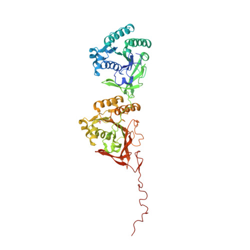
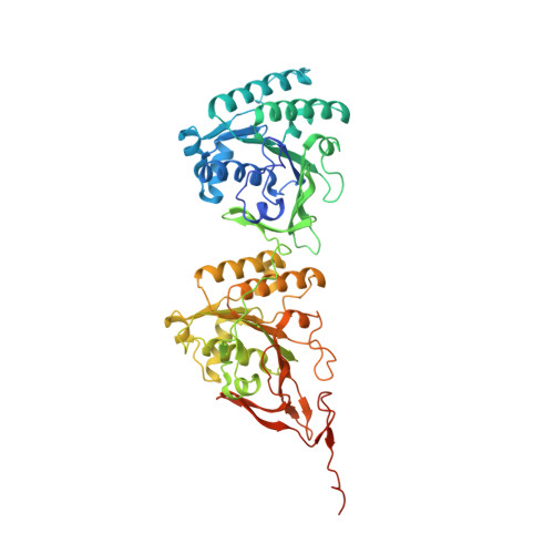
    3S1A

  • PubMed Abstract: 

    The circadian clock in the cyanobacterium Synechococcus elongatus is composed of a post-translational oscillator (PTO) that can be reconstituted in vitro from three different proteins in the presence of ATP and a transcription-translation feedback loop (TTFL). The homo-hexameric KaiC kinase, phosphatase and ATPase alternates between hypo- and hyper-phosphorylated states over the 24-h cycle, with KaiA enhancing phosphorylation, and KaiB antagonizing KaiA and promoting KaiC subunit exchange. SasA is a His kinase that relays output signals from the PTO formed by the three Kai proteins to the TTFL. Although the crystal structures for all three Kai proteins are known, atomic resolution structures of Kai and Kai/SasA protein complexes have remained elusive. Here, we present models of the KaiAC and KaiBC complexes derived from solution small angle X-ray scattering (SAXS), which are consistent with previous EM based models. We also present a combined SAXS/EM model of the KaiC/SasA complex, which has two N-terminal SasA sensory domains occupying positions on the C-terminal KaiC ring reminiscent of the orientations adopted by KaiB dimers. Using EM we demonstrate that KaiB and SasA compete for similar binding sites on KaiC. We also propose an EM based model of the ternary KaiABC complex that is consistent with the sequestering of KaiA by KaiB on KaiC during the PTO dephosphorylation phase. This work provides the first 3D-catalogue of protein-protein interactions in the KaiABC PTO and the output pathway mediated by SasA.


  • Organizational Affiliation

    Department of Biochemistry, School of Medicine, Vanderbilt University, Nashville, Tennessee, United States of America.


Macromolecules
Find similar proteins by:  (by identity cutoff)  |  3D Structure
Entity ID: 1
MoleculeChains Sequence LengthOrganismDetailsImage
Circadian clock protein kinase kaiC
A, F
525Synechococcus elongatus PCC 7942 = FACHB-805Mutation(s): 2 
Gene Names: kaiCsee0011Synpcc7942_1216
EC: 2.7.11.1
UniProt
Find proteins for Q79PF4 (Synechococcus elongatus (strain ATCC 33912 / PCC 7942 / FACHB-805))
Explore Q79PF4 
Go to UniProtKB:  Q79PF4
Entity Groups  
Sequence Clusters30% Identity50% Identity70% Identity90% Identity95% Identity100% Identity
UniProt GroupQ79PF4
Sequence Annotations
Expand
  • Reference Sequence
Find similar proteins by:  (by identity cutoff)  |  3D Structure
Entity ID: 2
MoleculeChains Sequence LengthOrganismDetailsImage
Circadian clock protein kinase kaiC
B, C, D, E
525Synechococcus elongatus PCC 7942 = FACHB-805Mutation(s): 2 
Gene Names: kaiCsee0011Synpcc7942_1216
EC: 2.7.11.1
UniProt
Find proteins for Q79PF4 (Synechococcus elongatus (strain ATCC 33912 / PCC 7942 / FACHB-805))
Explore Q79PF4 
Go to UniProtKB:  Q79PF4
Entity Groups  
Sequence Clusters30% Identity50% Identity70% Identity90% Identity95% Identity100% Identity
UniProt GroupQ79PF4
Sequence Annotations
Expand
  • Reference Sequence
Small Molecules
Ligands 2 Unique
IDChains Name / Formula / InChI Key2D Diagram3D Interactions
ATP
Query on ATP

Download Ideal Coordinates CCD File 
EA [auth E]
FA [auth E]
G [auth A]
H [auth A]
IA [auth F]
EA [auth E],
FA [auth E],
G [auth A],
H [auth A],
IA [auth F],
JA [auth F],
N [auth B],
O [auth B],
S [auth C],
T [auth C],
Y [auth D],
Z [auth D]
ADENOSINE-5'-TRIPHOSPHATE
C10 H16 N5 O13 P3
ZKHQWZAMYRWXGA-KQYNXXCUSA-N
MG
Query on MG

Download Ideal Coordinates CCD File 
AA [auth D]
BA [auth D]
CA [auth D]
DA [auth D]
GA [auth E]
AA [auth D],
BA [auth D],
CA [auth D],
DA [auth D],
GA [auth E],
HA [auth E],
I [auth A],
J [auth A],
K [auth A],
KA [auth F],
L [auth A],
LA [auth F],
M [auth B],
MA [auth F],
P [auth B],
Q [auth B],
R [auth B],
U [auth C],
V [auth C],
W [auth C],
X [auth C]
MAGNESIUM ION
Mg
JLVVSXFLKOJNIY-UHFFFAOYSA-N
Modified Residues  1 Unique
IDChains TypeFormula2D DiagramParent
SEP
Query on SEP
A, F
L-PEPTIDE LINKINGC3 H8 N O6 PSER
Experimental Data & Validation

Experimental Data

  • Method: X-RAY DIFFRACTION
  • Resolution: 3.00 Å
  • R-Value Free: 0.288 
  • R-Value Work: 0.242 
  • R-Value Observed: 0.268 
  • Space Group: P 21 21 21
Unit Cell:
Length ( Å )Angle ( ˚ )
a = 132.666α = 90
b = 135.494β = 90
c = 204.603γ = 90
Software Package:
Software NamePurpose
HKL-2000data collection
CNSrefinement
HKL-2000data reduction
HKL-2000data scaling
CNSphasing

Structure Validation

View Full Validation Report


Ligand Structure Quality Assessment 


Entry History 

Deposition Data

Revision History  (Full details and data files)

  • Version 1.0: 2011-09-21
    Type: Initial release
  • Version 1.1: 2023-09-13
    Changes: Data collection, Database references, Derived calculations, Refinement description