3RV1

Crystal structure of the N-terminal and RNase III domains of K. polysporus Dcr1 E224Q mutant


Experimental Data Snapshot

  • Method: X-RAY DIFFRACTION
  • Resolution: 1.98 Å
  • R-Value Free: 0.236 
  • R-Value Work: 0.194 
  • R-Value Observed: 0.197 

wwPDB Validation   3D Report Full Report


This is version 1.2 of the entry. See complete history


Literature

The inside-out mechanism of dicers from budding yeasts.

Weinberg, D.E.Nakanishi, K.Patel, D.J.Bartel, D.P.

(2011) Cell 146: 262-276

  • DOI: https://doi.org/10.1016/j.cell.2011.06.021
  • Primary Citation of Related Structures:  
    3RV0, 3RV1

  • PubMed Abstract: 

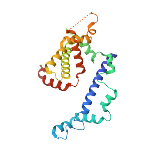
    The Dicer ribonuclease III (RNase III) enzymes process long double-stranded RNA (dsRNA) into small interfering RNAs (siRNAs) that direct RNA interference. Here, we describe the structure and activity of a catalytically active fragment of Kluyveromyces polysporus Dcr1, which represents the noncanonical Dicers found in budding yeasts. The crystal structure revealed a homodimer resembling that of bacterial RNase III but extended by a unique N-terminal domain, and it identified additional catalytic residues conserved throughout eukaryotic RNase III enzymes. Biochemical analyses showed that Dcr1 dimers bind cooperatively along the dsRNA substrate such that the distance between consecutive active sites determines the length of the siRNA products. Thus, unlike canonical Dicers, which successively remove siRNA duplexes from the dsRNA termini, budding-yeast Dicers initiate processing in the interior and work outward. The distinct mechanism of budding-yeast Dicers establishes a paradigm for natural molecular rulers and imparts substrate preferences with ramifications for biological function.


  • Organizational Affiliation

    Whitehead Institute for Biomedical Research, 9 Cambridge Center, Cambridge, MA 02142, USA.


Macromolecules
Find similar proteins by:  (by identity cutoff)  |  3D Structure
Entity ID: 1
MoleculeChains Sequence LengthOrganismDetailsImage
K. polysporus Dcr1
A, B
246Vanderwaltozyma polyspora DSM 70294Mutation(s): 1 
Gene Names: Kpol_455p11
UniProt
Find proteins for A7TR32 (Vanderwaltozyma polyspora (strain ATCC 22028 / DSM 70294 / BCRC 21397 / CBS 2163 / NBRC 10782 / NRRL Y-8283 / UCD 57-17))
Explore A7TR32 
Go to UniProtKB:  A7TR32
Entity Groups  
Sequence Clusters30% Identity50% Identity70% Identity90% Identity95% Identity100% Identity
UniProt GroupA7TR32
Sequence Annotations
Expand
  • Reference Sequence
Experimental Data & Validation

Experimental Data

  • Method: X-RAY DIFFRACTION
  • Resolution: 1.98 Å
  • R-Value Free: 0.236 
  • R-Value Work: 0.194 
  • R-Value Observed: 0.197 
  • Space Group: P 21 21 21
Unit Cell:
Length ( Å )Angle ( ˚ )
a = 58.907α = 90
b = 96.405β = 90
c = 101.449γ = 90
Software Package:
Software NamePurpose
DENZOdata reduction
SCALEPACKdata scaling
MOLREPphasing
PHENIXrefinement
PDB_EXTRACTdata extraction
HKL-2000data collection
HKL-2000data reduction
HKL-2000data scaling

Structure Validation

View Full Validation Report


Entry History 

Deposition Data

Revision History  (Full details and data files)

  • Version 1.0: 2011-08-03
    Type: Initial release
  • Version 1.1: 2017-11-08
    Changes: Refinement description
  • Version 1.2: 2024-02-28
    Changes: Data collection, Database references