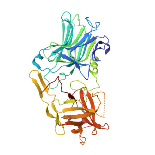
3RSJ

Structure of HCRF in complex with Ganglioside GD1a


Experimental Data Snapshot

  • Method: X-RAY DIFFRACTION
  • Resolution: 2.00 Å
  • R-Value Free: 0.263 
  • R-Value Work: 0.232 
  • R-Value Observed: 0.232 

wwPDB Validation   3D Report Full Report


This is version 2.1 of the entry. See complete history


Literature

Unique ganglioside recognition strategies for clostridial neurotoxins.

Benson, M.A.Fu, Z.Kim, J.J.Baldwin, M.R.

(2011) J Biol Chem 286: 34015-34022

  • DOI: https://doi.org/10.1074/jbc.M111.272054
  • Primary Citation of Related Structures:  
    3RSJ

  • PubMed Abstract: 

    Botulinum neurotoxins (BoNTs) and tetanus neurotoxin are the causative agents of the paralytic diseases botulism and tetanus, respectively. The potency of the clostridial neurotoxins (CNTs) relies primarily on their highly specific binding to nerve terminals and cleavage of SNARE proteins. Although individual CNTs utilize distinct proteins for entry, they share common ganglioside co-receptors. Here, we report the crystal structure of the BoNT/F receptor-binding domain in complex with the sugar moiety of ganglioside GD1a. GD1a binds in a shallow groove formed by the conserved peptide motif E … H … SXWY … G, with additional stabilizing interactions provided by two arginine residues. Comparative analysis of BoNT/F with other CNTs revealed several differences in the interactions of each toxin with ganglioside. Notably, exchange of BoNT/F His-1241 with the corresponding lysine residue of BoNT/E resulted in increased affinity for GD1a and conferred the ability to bind ganglioside GM1a. Conversely, BoNT/E was not able to bind GM1a, demonstrating a discrete mechanism of ganglioside recognition. These findings provide a structural basis for ganglioside binding among the CNTs and show that individual toxins utilize unique ganglioside recognition strategies.


  • Organizational Affiliation

    Department of Molecular Microbiology and Immunology, University of Missouri, Columbia, Missouri 65212, USA.


Macromolecules
Find similar proteins by:  (by identity cutoff)  |  3D Structure
Entity ID: 1
MoleculeChains Sequence LengthOrganismDetailsImage
BoNT/F
A, B, C, D
413Clostridium botulinumMutation(s): 0 
Gene Names: bont/fBotulinum Neurotoxin
UniProt
Find proteins for A7GBG3 (Clostridium botulinum (strain Langeland / NCTC 10281 / Type F))
Explore A7GBG3 
Go to UniProtKB:  A7GBG3
Entity Groups  
Sequence Clusters30% Identity50% Identity70% Identity90% Identity95% Identity100% Identity
UniProt GroupA7GBG3
Sequence Annotations
Expand
  • Reference Sequence
Oligosaccharides

Help

Entity ID: 2
MoleculeChains Length2D Diagram Glycosylation3D Interactions
N-acetyl-alpha-neuraminic acid-(2-3)-beta-D-galactopyranose-(1-3)-2-acetamido-2-deoxy-beta-D-galactopyranose
E, H
3N/A
Glycosylation Resources
GlyTouCan:  G02684WR
GlyCosmos:  G02684WR
GlyGen:  G02684WR
Entity ID: 3
MoleculeChains Length2D Diagram Glycosylation3D Interactions
N-acetyl-alpha-neuraminic acid-(2-3)-beta-D-galactopyranose-(1-3)-2-acetamido-2-deoxy-beta-D-galactopyranose-(1-4)-[N-acetyl-alpha-neuraminic acid-(2-3)]beta-D-galactopyranose
F
5N/A
Glycosylation Resources
GlyTouCan:  G78641QJ
GlyCosmos:  G78641QJ
GlyGen:  G78641QJ
Entity ID: 4
MoleculeChains Length2D Diagram Glycosylation3D Interactions
N-acetyl-alpha-neuraminic acid-(2-3)-beta-D-galactopyranose-(1-3)-2-acetamido-2-deoxy-beta-D-galactopyranose-(1-4)-beta-D-galactopyranose
G
4N/A
Glycosylation Resources
GlyTouCan:  G02162KR
GlyCosmos:  G02162KR
GlyGen:  G02162KR
Experimental Data & Validation

Experimental Data

  • Method: X-RAY DIFFRACTION
  • Resolution: 2.00 Å
  • R-Value Free: 0.263 
  • R-Value Work: 0.232 
  • R-Value Observed: 0.232 
  • Space Group: P 1
Unit Cell:
Length ( Å )Angle ( ˚ )
a = 65.468α = 72.55
b = 84.266β = 87.01
c = 117.6γ = 67.13
Software Package:
Software NamePurpose
ADSCdata collection
MOLREPphasing
CNSrefinement
HKL-3000data reduction
HKL-3000data scaling

Structure Validation

View Full Validation Report


Entry History 

Deposition Data

Revision History  (Full details and data files)

  • Version 1.0: 2011-08-17
    Type: Initial release
  • Version 1.1: 2011-08-31
    Changes: Database references
  • Version 1.2: 2011-10-12
    Changes: Database references
  • Version 1.3: 2018-01-24
    Changes: Structure summary
  • Version 2.0: 2020-07-29
    Type: Remediation
    Reason: Carbohydrate remediation
    Changes: Atomic model, Data collection, Derived calculations, Structure summary
  • Version 2.1: 2023-09-13
    Changes: Data collection, Database references, Refinement description, Structure summary