


Structural characterisation of staphylococcus aureus biotin protein ligase
Pendini, N.R., Yap, M.Y., Polyak, S.W., Cowieson, N., Traore, D.A.K., Booker, G.W., Wallace, J.C., Abell, A., Wilce, J.A., Wilce, M.C.J.To be published.
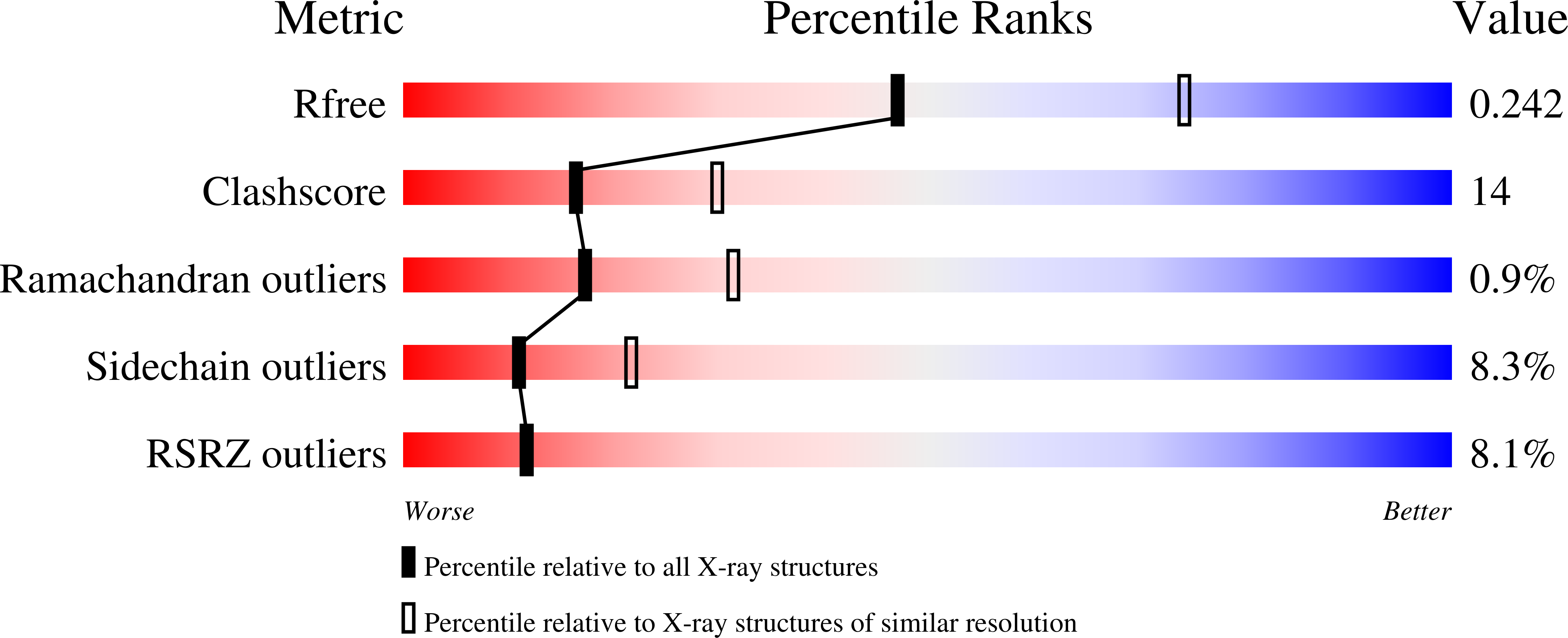
Experimental Data Snapshot
Entity ID: 1 | |||||
|---|---|---|---|---|---|
| Molecule | Chains | Sequence Length | Organism | Details | Image |
| Biotin-[acetyl-CoA-carboxylase] ligase | 323 | Staphylococcus aureus subsp. aureus ECT-R 2 | Mutation(s): 0 Gene Names: ECTR2_1310 EC: 6.3.4.15 | 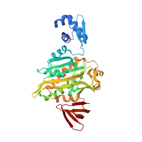 | |
Entity Groups | |||||
| Sequence Clusters | 30% Identity50% Identity70% Identity90% Identity95% Identity100% Identity | ||||
Sequence AnnotationsExpand | |||||
| |||||
| Ligands 1 Unique | |||||
|---|---|---|---|---|---|
| ID | Chains | Name / Formula / InChI Key | 2D Diagram | 3D Interactions | |
| BT5 Query on BT5 | B [auth A] | BIOTINYL-5-AMP C20 H28 N7 O9 P S UTQCSTJVMLODHM-RHCAYAJFSA-N |  | ||
| Length ( Å ) | Angle ( ˚ ) |
|---|---|
| a = 94.598 | α = 90 |
| b = 94.598 | β = 90 |
| c = 130.626 | γ = 90 |
| Software Name | Purpose |
|---|---|
| Blu-Ice | data collection |
| PHENIX | refinement |
| MOSFLM | data reduction |
| SCALA | data scaling |
RCSB PDB (citation) is hosted by
RCSB PDB is a member of the

