3RH8

Crystal Structure of the Light-state Dimer of Fungal Blue-Light Photoreceptor Vivid


Experimental Data Snapshot

  • Method: X-RAY DIFFRACTION
  • Resolution: 2.75 Å
  • R-Value Free: 0.287 
  • R-Value Work: 0.226 

wwPDB Validation   3D Report Full Report


Ligand Structure Quality Assessment 


This is version 1.2 of the entry. See complete history


Literature

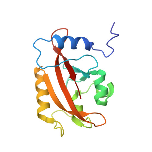
Structure of a Light-Activated LOV Protein Dimer That Regulates Transcription.

Vaidya, A.T.Chen, C.H.Dunlap, J.C.Loros, J.J.Crane, B.R.

(2011) Sci Signal 4: ra50-ra50

  • DOI: https://doi.org/10.1126/scisignal.2001945
  • Primary Citation of Related Structures:  
    3RH8

  • PubMed Abstract: 

    Light, oxygen, or voltage (LOV) protein domains are present in many signaling proteins in bacteria, archaea, protists, plants, and fungi. The LOV protein VIVID (VVD) of the filamentous fungus Neurospora crassa enables the organism to adapt to constant or increasing amounts of light and facilitates proper entrainment of circadian rhythms. Here, we determined the crystal structure of the fully light-adapted VVD dimer and reveal the mechanism by which light-driven conformational change alters the oligomeric state of the protein. Light-induced formation of a cysteinyl-flavin adduct generated a new hydrogen bond network that released the amino (N) terminus from the protein core and restructured an acceptor pocket for binding of the N terminus on the opposite subunit of the dimer. Substitution of residues critical for the switch between the monomeric and the dimeric states of the protein had profound effects on light adaptation in Neurospora. The mechanism of dimerization of VVD provides molecular details that explain how members of a large family of photoreceptors convert light responses to alterations in protein-protein interactions.


  • Organizational Affiliation

    Department of Chemistry and Chemical Biology, Cornell University, Ithaca, NY 14853, USA.


Macromolecules
Find similar proteins by:  (by identity cutoff)  |  3D Structure
Entity ID: 1
MoleculeChains Sequence LengthOrganismDetailsImage
Vivid PAS protein VVDA [auth B],
B [auth D]
148Neurospora crassaMutation(s): 0 
Gene Names: vvdG17A4.050
UniProt
Find proteins for Q9C3Y6 (Neurospora crassa)
Explore Q9C3Y6 
Go to UniProtKB:  Q9C3Y6
Entity Groups  
Sequence Clusters30% Identity50% Identity70% Identity90% Identity95% Identity100% Identity
UniProt GroupQ9C3Y6
Sequence Annotations
Expand
  • Reference Sequence
Small Molecules
Ligands 1 Unique
IDChains Name / Formula / InChI Key2D Diagram3D Interactions
FAD
Query on FAD

Download Ideal Coordinates CCD File 
C [auth B],
D
FLAVIN-ADENINE DINUCLEOTIDE
C27 H33 N9 O15 P2
VWWQXMAJTJZDQX-UYBVJOGSSA-N
Experimental Data & Validation

Experimental Data

  • Method: X-RAY DIFFRACTION
  • Resolution: 2.75 Å
  • R-Value Free: 0.287 
  • R-Value Work: 0.226 
  • Space Group: P 1 21 1
Unit Cell:
Length ( Å )Angle ( ˚ )
a = 37.647α = 90
b = 77.473β = 99.77
c = 54.696γ = 90
Software Package:
Software NamePurpose
DENZOdata reduction
SCALEPACKdata scaling
CNSrefinement
PDB_EXTRACTdata extraction
ADSCdata collection
PHASERphasing

Structure Validation

View Full Validation Report


Ligand Structure Quality Assessment 


Entry History 

Deposition Data

Revision History  (Full details and data files)

  • Version 1.0: 2011-09-21
    Type: Initial release
  • Version 1.1: 2017-11-08
    Changes: Refinement description
  • Version 1.2: 2023-09-13
    Changes: Data collection, Database references, Derived calculations, Refinement description