3RGU

Structure of Fap-NRa at pH 5.0


Experimental Data Snapshot

  • Method: X-RAY DIFFRACTION
  • Resolution: 3.00 Å
  • R-Value Free: 0.242 
  • R-Value Work: 0.223 
  • R-Value Observed: 0.224 

wwPDB Validation   3D Report Full Report


Ligand Structure Quality Assessment 


This is version 1.3 of the entry. See complete history


Literature

Structural insight into the role of Streptococcus parasanguinis Fap1 within oral biofilm formation.

Garnett, J.A.Simpson, P.J.Taylor, J.Benjamin, S.V.Tagliaferri, C.Cota, E.Chen, Y.Y.Wu, H.Matthews, S.

(2012) Biochem Biophys Res Commun 417: 421-426

  • DOI: https://doi.org/10.1016/j.bbrc.2011.11.131
  • Primary Citation of Related Structures:  
    3RGU

  • PubMed Abstract: 

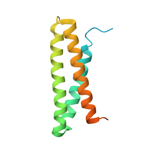
    The fimbriae-associated protein 1 (Fap1) is a major adhesin of Streptococcus parasanguinis, a primary colonizer of the oral cavity that plays an important role in the formation of dental plaque. Fap1 is an extracellular adhesive surface fibre belonging to the serine-rich repeat protein (SRRP) family, which plays a central role in the pathogenesis of streptococci and staphylococci. The N-terminal adhesive region of Fap1 (Fap1-NR) is composed of two domains (Fap1-NR(α) and Fap1-NR(β)) and is projected away from the bacterial surface via the extensive serine-rich repeat region, for adhesion to the salivary pellicle. The adhesive properties of Fap1 are modulated through a pH switch in which a reduction in pH results in a rearrangement between the Fap1-NR(α) and Fap1-NR(β) domains, which assists in the survival of S. parasanguinis in acidic environments. We have solved the structure of Fap1-NR(α) at pH 5.0 at 3.0Ǻ resolution and reveal how subtle rearrangements of the 3-helix bundle combined with a change in electrostatic potential mediates 'opening' and activation of the adhesive region. Further, we show that pH-dependent changes are critical for biofilm formation and present an atomic model for the inter-Fap1-NR interactions which have been assigned an important role in the biofilm formation.


  • Organizational Affiliation

    Department of Biological Sciences, Centre for Structural Biology, Imperial College London, London, UK.


Macromolecules
Find similar proteins by:  (by identity cutoff)  |  3D Structure
Entity ID: 1
MoleculeChains Sequence LengthOrganismDetailsImage
Fimbriae-associated protein Fap1
A, B, C, D
132Streptococcus parasanguinisMutation(s): 0 
Gene Names: fap1
UniProt
Find proteins for A1C3L3 (Streptococcus parasanguinis)
Explore A1C3L3 
Go to UniProtKB:  A1C3L3
Entity Groups  
Sequence Clusters30% Identity50% Identity70% Identity90% Identity95% Identity100% Identity
UniProt GroupA1C3L3
Sequence Annotations
Expand
  • Reference Sequence
Small Molecules
Ligands 1 Unique
IDChains Name / Formula / InChI Key2D Diagram3D Interactions
GLC
Query on GLC

Download Ideal Coordinates CCD File 
E [auth C]alpha-D-glucopyranose
C6 H12 O6
WQZGKKKJIJFFOK-DVKNGEFBSA-N
Experimental Data & Validation

Experimental Data

  • Method: X-RAY DIFFRACTION
  • Resolution: 3.00 Å
  • R-Value Free: 0.242 
  • R-Value Work: 0.223 
  • R-Value Observed: 0.224 
  • Space Group: P 43 21 2
Unit Cell:
Length ( Å )Angle ( ˚ )
a = 121.885α = 90
b = 121.885β = 90
c = 117.98γ = 90
Software Package:
Software NamePurpose
PHASERphasing
REFMACrefinement
MOSFLMdata reduction
SCALAdata scaling

Structure Validation

View Full Validation Report


Ligand Structure Quality Assessment 


Entry History 

Deposition Data

Revision History  (Full details and data files)

  • Version 1.0: 2011-12-28
    Type: Initial release
  • Version 1.1: 2012-02-01
    Changes: Database references
  • Version 1.2: 2020-07-29
    Type: Remediation
    Reason: Carbohydrate remediation
    Changes: Data collection, Database references, Derived calculations, Structure summary
  • Version 1.3: 2023-09-13
    Changes: Data collection, Database references, Refinement description, Structure summary