3RGA

Crystal structure of epoxide hydrolase for polyether lasalocid A biosynthesis


Experimental Data Snapshot

  • Method: X-RAY DIFFRACTION
  • Resolution: 1.59 Å
  • R-Value Free: 0.192 
  • R-Value Work: 0.173 
  • R-Value Observed: 0.174 

wwPDB Validation   3D Report Full Report


Ligand Structure Quality Assessment 


This is version 1.2 of the entry. See complete history


Literature

Enzymatic catalysis of anti-Baldwin ring closure in polyether biosynthesis

Hotta, K.Chen, X.Paton, R.S.Minami, A.Li, H.Swaminathan, K.Mathews, I.I.Watanabe, K.Oikawa, H.Houk, K.N.Kim, C.-Y.

(2012) Nature 483: 355-358

  • DOI: https://doi.org/10.1038/nature10865
  • Primary Citation of Related Structures:  
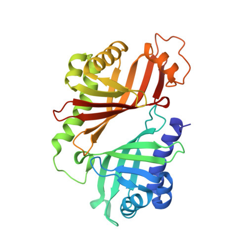
    3RGA

  • PubMed Abstract: 

    Polycyclic polyether natural products have fascinated chemists and biologists alike owing to their useful biological activity, highly complex structure and intriguing biosynthetic mechanisms. Following the original proposal for the polyepoxide origin of lasalocid and isolasalocid and the experimental determination of the origins of the oxygen and carbon atoms of both lasalocid and monensin, a unified stereochemical model for the biosynthesis of polyether ionophore antibiotics was proposed. The model was based on a cascade of nucleophilic ring closures of postulated polyepoxide substrates generated by stereospecific oxidation of all-trans polyene polyketide intermediates. Shortly thereafter, a related model was proposed for the biogenesis of marine ladder toxins, involving a series of nominally disfavoured anti-Baldwin, endo-tet epoxide-ring-opening reactions. Recently, we identified Lsd19 from the Streptomyces lasaliensis gene cluster as the epoxide hydrolase responsible for the epoxide-opening cyclization of bisepoxyprelasalocid A to form lasalocid A. Here we report the X-ray crystal structure of Lsd19 in complex with its substrate and product analogue to provide the first atomic structure-to our knowledge-of a natural enzyme capable of catalysing the disfavoured epoxide-opening cyclic ether formation. On the basis of our structural and computational studies, we propose a general mechanism for the enzymatic catalysis of polyether natural product biosynthesis.


  • Organizational Affiliation

    National University of Singapore, Department of Biological Sciences, 14 Science Drive 4, 117543 Singapore.


Macromolecules
Find similar proteins by:  (by identity cutoff)  |  3D Structure
Entity ID: 1
MoleculeChains Sequence LengthOrganismDetailsImage
Epoxide hydrolase283Streptomyces lasalocidiMutation(s): 0 
Gene Names: lasBlsd19
EC: 3.3.2
UniProt
Find proteins for B6ZK72 (Streptomyces lasalocidi)
Explore B6ZK72 
Go to UniProtKB:  B6ZK72
Entity Groups  
Sequence Clusters30% Identity50% Identity70% Identity90% Identity95% Identity100% Identity
UniProt GroupB6ZK72
Sequence Annotations
Expand
  • Reference Sequence
Small Molecules
Ligands 7 Unique
IDChains Name / Formula / InChI Key2D Diagram3D Interactions
ILD
Query on ILD

Download Ideal Coordinates CCD File 
C [auth A](4R,5S)-3-[(2R)-2-{(2S,2'R,4S,5S,5'R)-2,5'-diethyl-5'-[(1S)-1-hydroxyethyl]-4-methyloctahydro-2,2'-bifuran-5-yl}butanoyl]-4-methyl-5-phenyl-1,3-oxazolidin-2-one
C29 H43 N O6
SYWOXCFBKCIFEM-VTYWIBJUSA-N
LSB
Query on LSB

Download Ideal Coordinates CCD File 
B [auth A](4R,5S)-3-((2R,3S,4S)-2-ethyl-5-((2R,3R)-2-ethyl-3-(2-((2R,3R)-2-ethyl-3-methyloxiran-2-yl)ethyl)oxiran-2-yl)-3-hydroxy-4-methylpentanoyl)-4-methyl-5-phenyloxazolidin-2-one
C29 H43 N O6
PLMSSXURLNIBST-CWZDORBJSA-N
GOL
Query on GOL

Download Ideal Coordinates CCD File 
D [auth A],
E [auth A]
GLYCEROL
C3 H8 O3
PEDCQBHIVMGVHV-UHFFFAOYSA-N
ACT
Query on ACT

Download Ideal Coordinates CCD File 
F [auth A],
G [auth A]
ACETATE ION
C2 H3 O2
QTBSBXVTEAMEQO-UHFFFAOYSA-M
FMT
Query on FMT

Download Ideal Coordinates CCD File 
J [auth A],
K [auth A],
L [auth A]
FORMIC ACID
C H2 O2
BDAGIHXWWSANSR-UHFFFAOYSA-N
MOH
Query on MOH

Download Ideal Coordinates CCD File 
H [auth A],
I [auth A]
METHANOL
C H4 O
OKKJLVBELUTLKV-UHFFFAOYSA-N
NA
Query on NA

Download Ideal Coordinates CCD File 
M [auth A]SODIUM ION
Na
FKNQFGJONOIPTF-UHFFFAOYSA-N
Experimental Data & Validation

Experimental Data

  • Method: X-RAY DIFFRACTION
  • Resolution: 1.59 Å
  • R-Value Free: 0.192 
  • R-Value Work: 0.173 
  • R-Value Observed: 0.174 
  • Space Group: C 1 2 1
Unit Cell:
Length ( Å )Angle ( ˚ )
a = 117.85α = 90
b = 47.48β = 120.11
c = 62.33γ = 90
Software Package:
Software NamePurpose
Blu-Icedata collection
MOLREPphasing
REFMACrefinement
XDSdata reduction
XSCALEdata scaling

Structure Validation

View Full Validation Report


Ligand Structure Quality Assessment 


Entry History 

Deposition Data

Revision History  (Full details and data files)

  • Version 1.0: 2012-03-07
    Type: Initial release
  • Version 1.1: 2012-03-28
    Changes: Database references
  • Version 1.2: 2023-11-01
    Changes: Data collection, Database references, Derived calculations, Refinement description