3RB7

Crystal structure of CBD12 from CALX1.2


Experimental Data Snapshot

  • Method: X-RAY DIFFRACTION
  • Resolution: 2.90 Å
  • R-Value Free: 0.278 
  • R-Value Work: 0.231 

wwPDB Validation   3D Report Full Report


This is version 1.1 of the entry. See complete history


Literature

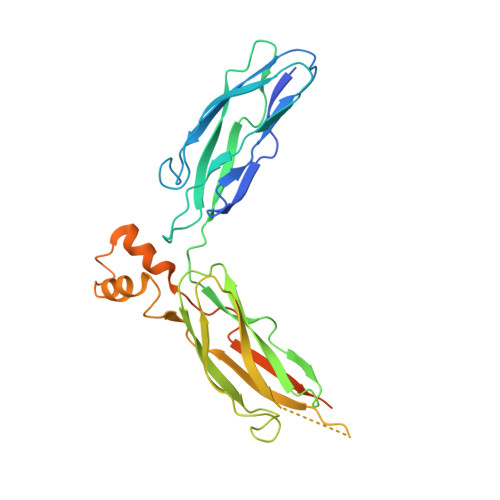
Structural Basis of the Ca(2+) Inhibitory Mechanism of Drosophila Na(+)/Ca(2+) Exchanger CALX and Its Modification by Alternative Splicing.

Wu, M.Tong, S.Gonzalez, J.Jayaraman, V.Spudich, J.L.Zheng, L.

(2011) Structure 19: 1509-1517

  • DOI: https://doi.org/10.1016/j.str.2011.07.008
  • Primary Citation of Related Structures:  
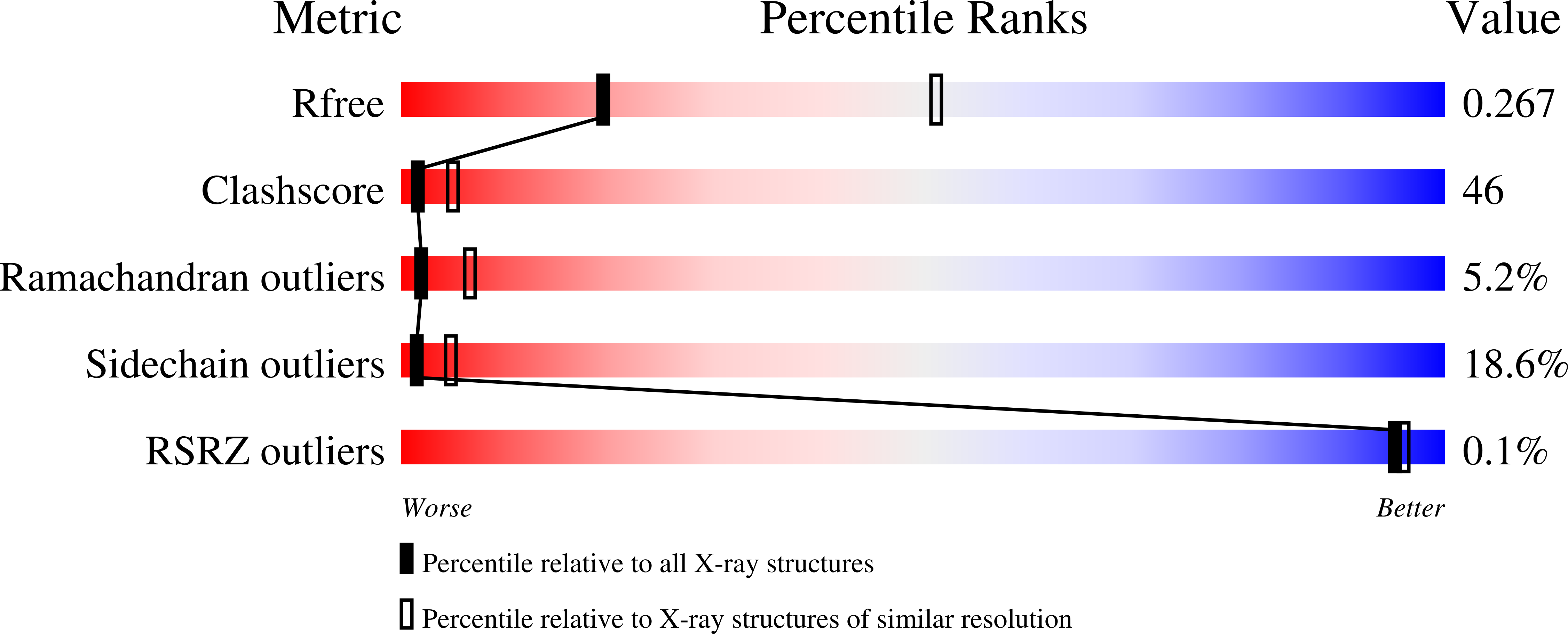
    3RB5, 3RB7

  • PubMed Abstract: 

    The Na(+)/Ca(2+) exchanger CALX promotes Ca(2+) efflux in Drosophila sensory neuronal cells to facilitate light-mediated Ca(2+) homeostasis. CALX activity is negatively regulated by specific Ca(2+) interaction within its two intracellular Ca(2+) regulatory domains CBD1 and CBD2, yet how the Ca(2+) binding is converted to molecular motion to operate the exchanger is unknown. Here, we report crystal structures of the entire Ca(2+) regulatory domain CBD12 from two alternative splicing isoforms, CALX 1.1 and 1.2, exhibiting distinct regulatory Ca(2+) dependency. The structures show an open V-shaped conformation with four Ca(2+) ions bound on the CBD domain interface, confirmed by LRET analysis. The structures together with Ca(2+)-binding analysis support that the Ca(2+) inhibition of CALX is achieved by interdomain conformational changes induced by Ca(2+) binding at CBD1. The conformational difference between the two isoforms also indicates that alternative splicing adjusts the interdomain orientation angle to modify the Ca(2+) regulatory property of the exchangers.


  • Organizational Affiliation

    Center for Membrane Biology, Department of Biochemistry and Molecular Biology, The University of Texas Houston Medical School, Houston, TX 77030, USA.


Macromolecules
Find similar proteins by:  (by identity cutoff)  |  3D Structure
Entity ID: 1
MoleculeChains Sequence LengthOrganismDetailsImage
Na/Ca exchange proteinA,
B,
C [auth E],
D [auth G]
298Drosophila melanogasterMutation(s): 0 
Gene Names: CalxNCXCG5685
UniProt
Find proteins for Q9VDG5 (Drosophila melanogaster)
Explore Q9VDG5 
Go to UniProtKB:  Q9VDG5
Entity Groups  
Sequence Clusters30% Identity50% Identity70% Identity90% Identity95% Identity100% Identity
UniProt GroupQ9VDG5
Sequence Annotations
Expand
  • Reference Sequence
Small Molecules
Ligands 2 Unique
IDChains Name / Formula / InChI Key2D Diagram3D Interactions
SO4
Query on SO4

Download Ideal Coordinates CCD File 
I [auth A],
J [auth B],
S [auth E],
X [auth G]
SULFATE ION
O4 S
QAOWNCQODCNURD-UHFFFAOYSA-L
CA
Query on CA

Download Ideal Coordinates CCD File 
E [auth A]
F [auth A]
G [auth A]
H [auth A]
K [auth B]
E [auth A],
F [auth A],
G [auth A],
H [auth A],
K [auth B],
L [auth B],
M [auth B],
N [auth B],
O [auth E],
P [auth E],
Q [auth E],
R [auth E],
T [auth G],
U [auth G],
V [auth G],
W [auth G]
CALCIUM ION
Ca
BHPQYMZQTOCNFJ-UHFFFAOYSA-N
Experimental Data & Validation

Experimental Data

  • Method: X-RAY DIFFRACTION
  • Resolution: 2.90 Å
  • R-Value Free: 0.278 
  • R-Value Work: 0.231 
  • Space Group: H 3
Unit Cell:
Length ( Å )Angle ( ˚ )
a = 107.535α = 90
b = 107.535β = 90
c = 358.65γ = 120
Software Package:
Software NamePurpose
CrystalCleardata collection
PHASERphasing
PHENIXrefinement
d*TREKdata reduction
d*TREKdata scaling

Structure Validation

View Full Validation Report


Entry History 

Deposition Data

Revision History  (Full details and data files)

  • Version 1.0: 2011-11-02
    Type: Initial release
  • Version 1.1: 2024-02-21
    Changes: Data collection, Database references, Derived calculations