3R7P

The crystal structure of I-LtrI


Experimental Data Snapshot

  • Method: X-RAY DIFFRACTION
  • Resolution: 2.70 Å
  • R-Value Free: 0.270 
  • R-Value Work: 0.192 
  • R-Value Observed: 0.196 

wwPDB Validation   3D Report Full Report


This is version 1.5 of the entry. See complete history


Literature

Tapping natural reservoirs of homing endonucleases for targeted gene modification.

Takeuchi, R.Lambert, A.R.Mak, A.N.Jacoby, K.Dickson, R.J.Gloor, G.B.Scharenberg, A.M.Edgell, D.R.Stoddard, B.L.

(2011) Proc Natl Acad Sci U S A 108: 13077-13082

  • DOI: https://doi.org/10.1073/pnas.1107719108
  • Primary Citation of Related Structures:  
    3QQY, 3R7P

  • PubMed Abstract: 

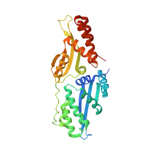
    Homing endonucleases mobilize their own genes by generating double-strand breaks at individual target sites within potential host DNA. Because of their high specificity, these proteins are used for "genome editing" in higher eukaryotes. However, alteration of homing endonuclease specificity is quite challenging. Here we describe the identification and phylogenetic analysis of over 200 naturally occurring LAGLIDADG homing endonucleases (LHEs). Biochemical and structural characterization of endonucleases from one clade within the phylogenetic tree demonstrates strong conservation of protein structure contrasted against highly diverged DNA target sites and indicates that a significant fraction of these proteins are sufficiently stable and active to serve as engineering scaffolds. This information was exploited to create a targeting enzyme to disrupt the endogenous monoamine oxidase B gene in human cells. The ubiquitous presence and diversity of LHEs described in this study may facilitate the creation of many tailored nucleases for genome editing.


  • Organizational Affiliation

    Division of Basic Sciences, Fred Hutchinson Cancer Research Center, 1100 Fairview Avenue North A3-025, Seattle, WA 98109, USA.


Macromolecules

Find similar proteins by:  (by identity cutoff)  |  3D Structure
Entity ID: 1
MoleculeChains Sequence LengthOrganismDetailsImage
Ribosomal protein 3/homing endonuclease-like fusion protein315Leptographium truncatumMutation(s): 0 
Gene Names: rps3/HEG fusion
EC: 3.1.21.1
UniProt
Find proteins for C7SWF3 (Leptographium truncatum)
Explore C7SWF3 
Go to UniProtKB:  C7SWF3
Entity Groups  
Sequence Clusters30% Identity50% Identity70% Identity90% Identity95% Identity100% Identity
UniProt GroupC7SWF3
Sequence Annotations
Expand
  • Reference Sequence

Find similar nucleic acids by:  Sequence   |   3D Structure  

Entity ID: 2
MoleculeChains LengthOrganismImage
DNA (5'-D(*GP*GP*TP*CP*TP*AP*AP*AP*CP*GP*TP*CP*GP*TP*AP*T)-3')16N/A
Sequence Annotations
Expand
  • Reference Sequence

Find similar nucleic acids by:  Sequence   |   3D Structure  

Entity ID: 3
MoleculeChains LengthOrganismImage
DNA (5'-D(P*AP*GP*GP*AP*GP*CP*AP*TP*TP*TP*G)-3')11N/A
Sequence Annotations
Expand
  • Reference Sequence

Find similar nucleic acids by:  Sequence   |   3D Structure  

Entity ID: 4
MoleculeChains LengthOrganismImage
DNA (5'-D(*CP*AP*AP*AP*TP*GP*CP*TP*CP*CP*TP*AP*TP*AP*C)-3')15N/A
Sequence Annotations
Expand
  • Reference Sequence

Find similar nucleic acids by:  Sequence   |   3D Structure  

Entity ID: 5
MoleculeChains LengthOrganismImage
DNA (5'-D(P*GP*AP*CP*GP*TP*TP*TP*AP*GP*AP*CP*C)-3')12N/A
Sequence Annotations
Expand
  • Reference Sequence
Experimental Data & Validation

Experimental Data

  • Method: X-RAY DIFFRACTION
  • Resolution: 2.70 Å
  • R-Value Free: 0.270 
  • R-Value Work: 0.192 
  • R-Value Observed: 0.196 
  • Space Group: C 1 2 1
Unit Cell:
Length ( Å )Angle ( ˚ )
a = 113.089α = 90
b = 42.595β = 110.63
c = 103.195γ = 90
Software Package:
Software NamePurpose
HKL-2000data collection
PHASESphasing
REFMACrefinement
HKL-2000data reduction
HKL-2000data scaling

Structure Validation

View Full Validation Report


Entry History 

Deposition Data

Revision History  (Full details and data files)

  • Version 1.0: 2011-07-20
    Type: Initial release
  • Version 1.1: 2011-08-10
    Changes: Database references
  • Version 1.2: 2011-08-24
    Changes: Database references
  • Version 1.3: 2013-04-03
    Changes: Structure summary
  • Version 1.4: 2015-01-21
    Changes: Database references
  • Version 1.5: 2023-09-13
    Changes: Data collection, Database references, Derived calculations, Refinement description