3R1V

Odorant Binding Protein 7 from Anopheles gambiae with Four Disulfide Bridges, in complex with an azo compound


Experimental Data Snapshot

  • Method: X-RAY DIFFRACTION
  • Resolution: 2.19 Å
  • R-Value Free: 0.255 
  • R-Value Work: 0.242 
  • R-Value Observed: 0.243 

wwPDB Validation   3D Report Full Report


Ligand Structure Quality Assessment 


This is version 1.2 of the entry. See complete history


Literature

The Crystal Structure of Odorant Binding Protein 7 from Anopheles gambiae Exhibits an Outstanding Adaptability of Its Binding Site.

Lagarde, A.Spinelli, S.Tegoni, M.He, X.Field, L.Zhou, J.J.Cambillau, C.

(2011) J Mol Biol 414: 401-412

  • DOI: https://doi.org/10.1016/j.jmb.2011.10.005
  • Primary Citation of Related Structures:  
    3R1O, 3R1P, 3R1V

  • PubMed Abstract: 

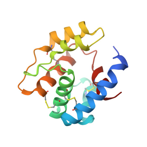
    Anopheles gambiae (Agam) targets human and animals by using its olfactory system, leading to the spread of Plasmodium falciparum, the malaria vector. Odorant binding proteins (OBPs) participate to the first event in odorant recognition and constitute an interesting target for insect control. OBPs interact with olfactory receptors to which they deliver the odorant molecule. We have undertaken a large-scale study of proteins belonging to the olfactory system of Agam with in mind of designing strong olfactory repellants. Here, we report the expression, three-dimensional structures and binding properties of AgamOBP07, a member of a new structural class of OBPs, characterized by the occurrence of eight cysteines. We showed that AgamOBP07 possesses seven α-helices and four disulfide bridges, instead of six α-helices and three disulfide bridges in classical OBPs. The extra seventh helix is located at the surface of the protein, locked by the fourth disulfide bridge, and forms a wall of the internal cavity. The binding site of the protein is mainly hydrophobic, elongated and open and is able to accommodate elongated ligands, linear or polycyclic, as suggested also by binding experiments. An elongated electron density was observed in the internal cavity of the purified protein, belonging to a serendipitous ligand. The structure of AgamOBP07 in complex with an azo-bicyclic model compound reveals that a large conformational change in the protein has reshaped its binding site, provoking helix 4 unfolding and doubling of the cavity volume.


  • Organizational Affiliation

    Architecture et Fonction des Macromolécules Biologiques, UMR 6098 CNRS and Universités of Marseille, 13288 Marseille cedex 9, France.


Macromolecules
Find similar proteins by:  (by identity cutoff)  |  3D Structure
Entity ID: 1
MoleculeChains Sequence LengthOrganismDetailsImage
Odorant binding protein, antennal
A, B
127Anopheles gambiaeMutation(s): 0 
Gene Names: OBP7AgamOBP7AGAP001556agCG57323
UniProt
Find proteins for Q7PXT9 (Anopheles gambiae)
Explore Q7PXT9 
Go to UniProtKB:  Q7PXT9
Entity Groups  
Sequence Clusters30% Identity50% Identity70% Identity90% Identity95% Identity100% Identity
UniProt GroupQ7PXT9
Sequence Annotations
Expand
  • Reference Sequence
Small Molecules
Ligands 1 Unique
IDChains Name / Formula / InChI Key2D Diagram3D Interactions
AZB
Query on AZB

Download Ideal Coordinates CCD File 
C [auth A],
D [auth B]
4-{(E)-[4-(propan-2-yl)phenyl]diazenyl}phenol
C15 H16 N2 O
NQJVTPMZGHHWGR-WUKNDPDISA-N
Binding Affinity Annotations 
IDSourceBinding Affinity
AZB Binding MOAD:  3R1V Kd: 120 (nM) from 1 assay(s)
PDBBind:  3R1V Kd: 120 (nM) from 1 assay(s)
Experimental Data & Validation

Experimental Data

  • Method: X-RAY DIFFRACTION
  • Resolution: 2.19 Å
  • R-Value Free: 0.255 
  • R-Value Work: 0.242 
  • R-Value Observed: 0.243 
  • Space Group: P 1 21 1
Unit Cell:
Length ( Å )Angle ( ˚ )
a = 48.76α = 90
b = 41.97β = 105.97
c = 57.94γ = 90
Software Package:
Software NamePurpose
ADSCdata collection
MOLREPphasing
BUSTERrefinement
XDSdata reduction
XSCALEdata scaling

Structure Validation

View Full Validation Report


Ligand Structure Quality Assessment 


Entry History 

Deposition Data

Revision History  (Full details and data files)

  • Version 1.0: 2011-10-19
    Type: Initial release
  • Version 1.1: 2011-11-09
    Changes: Database references
  • Version 1.2: 2011-12-21
    Changes: Database references