3QYQ

1.8 Angstrom resolution crystal structure of a putative deoxyribose-phosphate aldolase from Toxoplasma gondii ME49


Experimental Data Snapshot

  • Method: X-RAY DIFFRACTION
  • Resolution: 1.80 Å
  • R-Value Free: 0.201 
  • R-Value Work: 0.166 
  • R-Value Observed: 0.167 

wwPDB Validation   3D Report Full Report


This is version 2.1 of the entry. See complete history


Literature

Structural and functional divergence of the aldolase fold in Toxoplasma gondii.

Tonkin, M.L.Halavaty, A.S.Ramaswamy, R.Ruan, J.Igarashi, M.Ngo, H.M.Boulanger, M.J.

(2015) J Mol Biol 427: 840-852

  • DOI: https://doi.org/10.1016/j.jmb.2014.09.019
  • Primary Citation of Related Structures:  
    3QYQ, 4D2J, 4EIV

  • PubMed Abstract: 

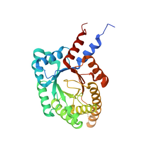
    Parasites of the phylum Apicomplexa are highly successful pathogens of humans and animals worldwide. As obligate intracellular parasites, they have significant energy requirements for invasion and gliding motility that are supplied by various metabolic pathways. Aldolases have emerged as key enzymes involved in these pathways, and all apicomplexans express one or both of fructose 1,6-bisphosphate (F16BP) aldolase and 2-deoxyribose 5-phosphate (dR5P) aldolase (DERA). Intriguingly, Toxoplasma gondii, a highly successful apicomplexan parasite, expresses F16BP aldolase (TgALD1), d5RP aldolase (TgDERA), and a divergent dR5P aldolase-like protein (TgDPA) exclusively in the latent bradyzoite stage. While the importance of TgALD1 in glycolysis is well established and TgDERA is also likely to be involved in parasite metabolism, the detailed function of TgDPA remains elusive. To gain mechanistic insight into the function of different T. gondii aldolases, we first determined the crystal structures of TgALD1 and TgDPA. Structural analysis revealed that both aldolases adopt a TIM barrel fold accessorized with divergent secondary structure elements. Structural comparison of TgALD1 and TgDPA with members of their respective enzyme families revealed that, while the active-site residues are conserved in TgALD1, key catalytic residues are absent in TgDPA. Consistent with this observation, biochemical assays showed that, while TgALD1 was active on F16BP, TgDPA was inactive on dR5P. Intriguingly, both aldolases are competent to bind polymerized actin in vitro. Altogether, structural and biochemical analyses of T. gondii aldolase and aldolase-like proteins reveal diverse functionalization of the classic TIM barrel aldolase fold.


  • Organizational Affiliation

    Biochemistry and Microbiology, University of Victoria, PO Box 3055 STN CSC, Victoria, BC, Canada V8W 3P6.


Macromolecules
Find similar proteins by:  (by identity cutoff)  |  3D Structure
Entity ID: 1
MoleculeChains Sequence LengthOrganismDetailsImage
Deoxyribose-phosphate aldolase, putative
A, B, C, D
293Toxoplasma gondii ME49Mutation(s): 0 
Gene Names: TGME49_118750
EC: 4.1.2.4
UniProt
Find proteins for B9PVB4 (Toxoplasma gondii (strain ATCC 50861 / VEG))
Explore B9PVB4 
Go to UniProtKB:  B9PVB4
Entity Groups  
Sequence Clusters30% Identity50% Identity70% Identity90% Identity95% Identity100% Identity
UniProt GroupB9PVB4
Sequence Annotations
Expand
  • Reference Sequence
Oligosaccharides

Help

Entity ID: 2
MoleculeChains Length2D Diagram Glycosylation3D Interactions
beta-D-fructofuranose-(2-1)-alpha-D-glucopyranose
E, F, G
2N/A
Glycosylation Resources
GlyTouCan:  G05551OP
GlyCosmos:  G05551OP
Small Molecules
Ligands 1 Unique
IDChains Name / Formula / InChI Key2D Diagram3D Interactions
SO4
Query on SO4

Download Ideal Coordinates CCD File 
H [auth A]
I [auth A]
J [auth A]
K [auth A]
L [auth A]
H [auth A],
I [auth A],
J [auth A],
K [auth A],
L [auth A],
M [auth A],
N [auth B],
O [auth B],
P [auth B],
Q [auth C],
R [auth C],
S [auth D],
T [auth D],
U [auth D],
V [auth D],
W [auth D]
SULFATE ION
O4 S
QAOWNCQODCNURD-UHFFFAOYSA-L
Biologically Interesting Molecules (External Reference) 1 Unique
Experimental Data & Validation

Experimental Data

  • Method: X-RAY DIFFRACTION
  • Resolution: 1.80 Å
  • R-Value Free: 0.201 
  • R-Value Work: 0.166 
  • R-Value Observed: 0.167 
  • Space Group: P 1
  • Diffraction Data: https://doi.org/10.18430/M33QYQ
Unit Cell:
Length ( Å )Angle ( ˚ )
a = 70.923α = 62.9
b = 72.194β = 89.8
c = 73.048γ = 88.85
Software Package:
Software NamePurpose
Blu-Icedata collection
PHASERphasing
REFMACrefinement
HKL-2000data reduction
HKL-2000data scaling

Structure Validation

View Full Validation Report


Entry History 

Revision History  (Full details and data files)

  • Version 1.0: 2011-03-30
    Type: Initial release
  • Version 1.1: 2011-07-13
    Changes: Version format compliance
  • Version 1.2: 2014-01-15
    Changes: Refinement description
  • Version 1.3: 2014-11-12
    Changes: Database references
  • Version 1.4: 2015-04-08
    Changes: Database references
  • Version 1.5: 2017-11-08
    Changes: Refinement description
  • Version 2.0: 2020-07-29
    Type: Remediation
    Reason: Carbohydrate remediation
    Changes: Atomic model, Data collection, Database references, Derived calculations, Non-polymer description, Structure summary
  • Version 2.1: 2023-09-13
    Changes: Data collection, Database references, Refinement description, Structure summary