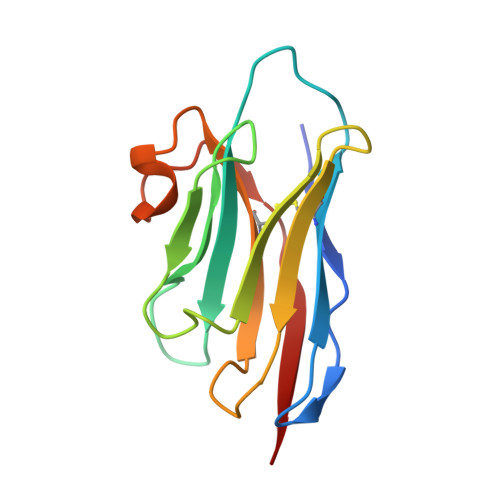
An anti-hapten camelid antibody reveals a cryptic binding site with significant energetic contributions from a nonhypervariable loop.
Fanning, S.W., Horn, J.R.(2011) Protein Sci 20: 1196-1207
- PubMed: 21557375
- DOI: https://doi.org/10.1002/pro.648
- Primary Citation of Related Structures:
3QXT, 3QXU, 3QXV, 3QXW - PubMed Abstract:
Conventional anti-hapten antibodies typically bind low-molecular weight compounds (haptens) in the crevice between the variable heavy and light chains. Conversely, heavy chain-only camelid antibodies, which lack a light chain, must rely entirely on a single variable domain to recognize haptens. While several anti-hapten VHHs have been generated, little is known regarding the underlying structural and thermodynamic basis for hapten recognition. Here, an anti-methotrexate VHH (anti-MTX VHH) was generated using grafting methods whereby the three complementarity determining regions (CDRs) were inserted onto an existing VHH framework. Thermodynamic analysis of the anti-MTX VHH CDR1-3 Graft revealed a micromolar binding affinity, while the crystal structure of the complex revealed a somewhat surprising noncanonical binding site which involved MTX tunneling under the CDR1 loop. Due to the close proximity of MTX to CDR4, a nonhypervariable loop, the CDR4 loop sequence was subsequently introduced into the CDR1-3 graft, which resulted in a dramatic 1000-fold increase in the binding affinity. Crystal structure analysis of both the free and complex anti-MTX CDR1-4 graft revealed CDR4 plays a significant role in both intermolecular contacts and binding site conformation that appear to contribute toward high affinity binding. Additionally, the anti-MTX VHH possessed relatively high specificity for MTX over closely related compounds aminopterin and folate, demonstrating that VHH domains are capable of binding low-molecular weight ligands with high affinity and specificity, despite their reduced interface.
Organizational Affiliation:
Department of Chemistry and Biochemistry, Northern Illinois University, DeKalb, IL 60115, USA.