Structural Basis for Inhibition of Cathepsin B Drug Target from the Human Blood Fluke, Schistosoma mansoni.
Jilkova, A., Rezacova, P., Lepsik, M., Horn, M., Vachova, J., Fanfrlik, J., Brynda, J., McKerrow, J.H., Caffrey, C.R., Mares, M.(2011) J Biol Chem 286: 35770-35781
- PubMed: 21832058
- DOI: https://doi.org/10.1074/jbc.M111.271304
- Primary Citation of Related Structures:
3QSD, 3S3Q, 3S3R - PubMed Abstract:
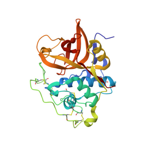
Schistosomiasis caused by a parasitic blood fluke of the genus Schistosoma afflicts over 200 million people worldwide. Schistosoma mansoni cathepsin B1 (SmCB1) is a gut-associated peptidase that digests host blood proteins as a source of nutrients. It is under investigation as a drug target. To further this goal, we report three crystal structures of SmCB1 complexed with peptidomimetic inhibitors as follows: the epoxide CA074 at 1.3 Å resolution and the vinyl sulfones K11017 and K11777 at 1.8 and 2.5 Å resolutions, respectively. Interactions of the inhibitors with the subsites of the active-site cleft were evaluated by quantum chemical calculations. These data and inhibition profiling with a panel of vinyl sulfone derivatives identify key binding interactions and provide insight into the specificity of SmCB1 inhibition. Furthermore, hydrolysis profiling of SmCB1 using synthetic peptides and the natural substrate hemoglobin revealed that carboxydipeptidase activity predominates over endopeptidolysis, thereby demonstrating the contribution of the occluding loop that restricts access to the active-site cleft. Critically, the severity of phenotypes induced in the parasite by vinyl sulfone inhibitors correlated with enzyme inhibition, providing support that SmCB1 is a valuable drug target. The present structure and inhibitor interaction data provide a footing for the rational design of anti-schistosomal inhibitors.
Organizational Affiliation:
Institute of Organic Chemistry and Biochemistry, Academy of Sciences of the Czech Republic, 16610 Prague, Czech Republic; Department of Biochemistry, Faculty of Science, Charles University, 12843 Prague, Czech Republic.