3QRC

The crystal structure of Ail, the attachment invasion locus protein of Yersinia pestis, in complex with the heparin analogue sucrose octasulfate


Experimental Data Snapshot

  • Method: X-RAY DIFFRACTION
  • Resolution: 1.85 Å
  • R-Value Free: 0.221 
  • R-Value Work: 0.204 
  • R-Value Observed: 0.204 

wwPDB Validation   3D Report Full Report


Ligand Structure Quality Assessment 


This is version 2.1 of the entry. See complete history


Literature

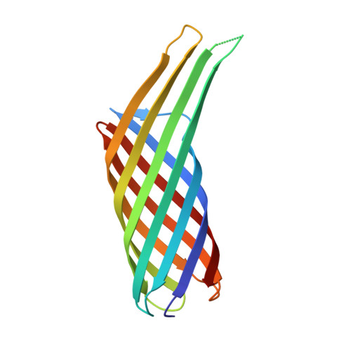
Structural Insights into Ail-Mediated Adhesion in Yersinia pestis.

Yamashita, S.Lukacik, P.Barnard, T.J.Noinaj, N.Felek, S.Tsang, T.M.Krukonis, E.S.Hinnebusch, B.J.Buchanan, S.K.

(2011) Structure 19: 1672-1682

  • DOI: https://doi.org/10.1016/j.str.2011.08.010
  • Primary Citation of Related Structures:  
    3QRA, 3QRC

  • PubMed Abstract: 

    Ail is an outer membrane protein from Yersinia pestis that is highly expressed in a rodent model of bubonic plague, making it a good candidate for vaccine development. Ail is important for attaching to host cells and evading host immune responses, facilitating rapid progression of a plague infection. Binding to host cells is important for injection of cytotoxic Yersinia outer proteins. To learn more about how Ail mediates adhesion, we solved two high-resolution crystal structures of Ail, with no ligand bound and in complex with a heparin analog called sucrose octasulfate. We identified multiple adhesion targets, including laminin and heparin, and showed that a 40 kDa domain of laminin called LG4-5 specifically binds to Ail. We also evaluated the contribution of laminin to delivery of Yops to HEp-2 cells. This work constitutes a structural description of how a bacterial outer membrane protein uses a multivalent approach to bind host cells.


  • Organizational Affiliation

    Laboratory of Molecular Biology, National Institute of Diabetes and Digestive and Kidney Diseases, National Institutes of Health, Bethesda, MD 20892-8030, USA.


Macromolecules
Find similar proteins by:  (by identity cutoff)  |  3D Structure
Entity ID: 1
MoleculeChains Sequence LengthOrganismDetailsImage
Attachment invasion locus protein
A, B
157Yersinia pestisMutation(s): 0 
Gene Names: ailYPO2905
Membrane Entity: Yes 
UniProt
Find proteins for Q8D0Z7 (Yersinia pestis)
Explore Q8D0Z7 
Go to UniProtKB:  Q8D0Z7
Entity Groups  
Sequence Clusters30% Identity50% Identity70% Identity90% Identity95% Identity100% Identity
UniProt GroupQ8D0Z7
Sequence Annotations
Expand
  • Reference Sequence
Oligosaccharides

Help

Entity ID: 2
MoleculeChains Length2D Diagram Glycosylation3D Interactions
1,3,4,6-tetra-O-sulfo-beta-D-fructofuranose-(2-1)-2,3,4,6-tetra-O-sulfonato-alpha-D-glucopyranose
C, D
2N/A
Glycosylation Resources
GlyTouCan:  G97555GV
GlyCosmos:  G97555GV
Biologically Interesting Molecules (External Reference) 1 Unique
Experimental Data & Validation

Experimental Data

  • Method: X-RAY DIFFRACTION
  • Resolution: 1.85 Å
  • R-Value Free: 0.221 
  • R-Value Work: 0.204 
  • R-Value Observed: 0.204 
  • Space Group: P 32
Unit Cell:
Length ( Å )Angle ( ˚ )
a = 91.382α = 90
b = 91.382β = 90
c = 47.496γ = 120
Software Package:
Software NamePurpose
SERGUIdata collection
PHASERphasing
PHENIXrefinement
HKL-2000data reduction
HKL-2000data scaling

Structure Validation

View Full Validation Report


Ligand Structure Quality Assessment 


Entry History 

Deposition Data

Revision History  (Full details and data files)

  • Version 1.0: 2011-11-23
    Type: Initial release
  • Version 2.0: 2020-07-29
    Type: Remediation
    Reason: Carbohydrate remediation
    Changes: Advisory, Atomic model, Data collection, Database references, Derived calculations, Non-polymer description, Structure summary
  • Version 2.1: 2023-09-13
    Changes: Data collection, Database references, Refinement description, Structure summary