3QR5

Structure of the first domain of a cardiac Ryanodine Receptor mutant with exon 3 deleted


Experimental Data Snapshot

  • Method: X-RAY DIFFRACTION
  • Resolution: 2.30 Å
  • R-Value Free: 0.250 
  • R-Value Work: 0.209 
  • R-Value Observed: 0.211 

wwPDB Validation   3D Report Full Report


This is version 1.4 of the entry. See complete history


Literature

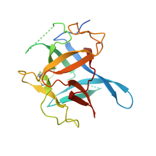
The deletion of exon 3 in the cardiac ryanodine receptor is rescued by beta strand switching.

Lobo, P.A.Kimlicka, L.Tung, C.C.Van Petegem, F.

(2011) Structure 19: 790-798

  • DOI: https://doi.org/10.1016/j.str.2011.03.016
  • Primary Citation of Related Structures:  
    3QR5

  • PubMed Abstract: 

    Mutations in the cardiac Ryanodine Receptor (RYR2) are linked to triggered arrhythmias. Removal of exon 3 results in a severe form of catecholaminergic polymorphic ventricular tachycardia (CPVT). This exon encodes secondary structure elements that are crucial for folding of the N-terminal domain (NTD), raising the question of why the deletion is neither lethal nor confers a loss of function. We determined the 2.3 Å crystal structure of the NTD lacking exon 3. The removal causes a structural rescue whereby a flexible loop inserts itself into the β trefoil domain and increases thermal stability. The exon 3 deletion is not tolerated in the corresponding RYR1 domain. The rescue shows a novel mechanism by which RYR2 channels can adjust their Ca²⁺ release properties through altering the structure of the NTD. Despite the rescue, the deletion affects interfaces with other RYR2 domains. We propose that relative movement of the NTD is allosterically coupled to the pore region.


  • Organizational Affiliation

    Department of Biochemistry and Molecular Biology, University of British Columbia, 2350 Health Sciences Mall, room 2.320, Vancouver, BC V6T1Z3, Canada.


Macromolecules
Find similar proteins by:  (by identity cutoff)  |  3D Structure
Entity ID: 1
MoleculeChains Sequence LengthOrganismDetailsImage
Cardiac Ca2+ release channel
A, B
185Mus musculusMutation(s): 0 
Gene Names: Ryr2
UniProt & NIH Common Fund Data Resources
Find proteins for E9Q401 (Mus musculus)
Explore E9Q401 
Go to UniProtKB:  E9Q401
IMPC:  MGI:99685
Entity Groups  
Sequence Clusters30% Identity50% Identity70% Identity90% Identity95% Identity100% Identity
UniProt GroupE9Q401
Sequence Annotations
Expand
  • Reference Sequence
Experimental Data & Validation

Experimental Data

  • Method: X-RAY DIFFRACTION
  • Resolution: 2.30 Å
  • R-Value Free: 0.250 
  • R-Value Work: 0.209 
  • R-Value Observed: 0.211 
  • Space Group: C 1 2 1
Unit Cell:
Length ( Å )Angle ( ˚ )
a = 95.37α = 90
b = 44.204β = 90.98
c = 76.606γ = 90
Software Package:
Software NamePurpose
SCALEPACKdata scaling
PHASERphasing
REFMACrefinement
PDB_EXTRACTdata extraction
HKL-2000data collection
DENZOdata reduction

Structure Validation

View Full Validation Report


Entry History 

Deposition Data

Revision History  (Full details and data files)

  • Version 1.0: 2011-06-08
    Type: Initial release
  • Version 1.1: 2011-07-13
    Changes: Version format compliance
  • Version 1.2: 2017-07-26
    Changes: Refinement description, Source and taxonomy
  • Version 1.3: 2017-11-08
    Changes: Refinement description
  • Version 1.4: 2023-07-26
    Changes: Database references