Characterization of dye-decolorizing peroxidases from Rhodococcus jostii RHA1.
Roberts, J.N., Singh, R., Grigg, J.C., Murphy, M.E., Bugg, T.D., Eltis, L.D.(2011) Biochemistry 50: 5108-5119
- PubMed: 21534572
- DOI: https://doi.org/10.1021/bi200427h
- Primary Citation of Related Structures:
3QNR, 3QNS - PubMed Abstract:
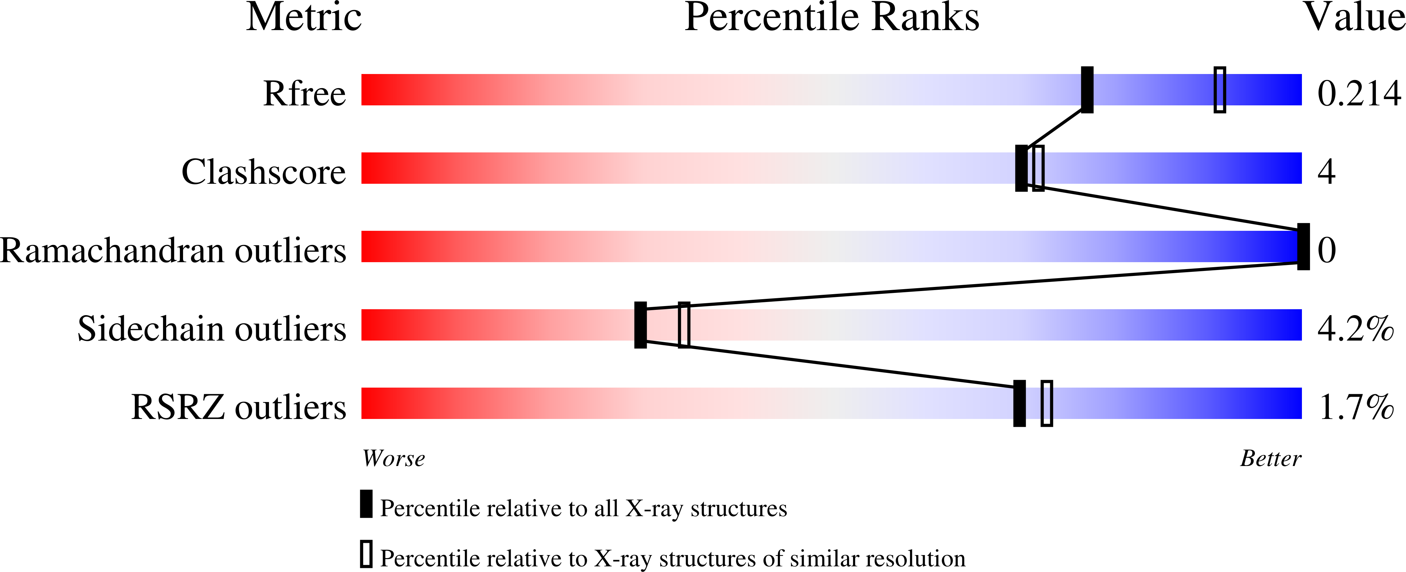
The soil bacterium Rhodococcus jostii RHA1 contains two dye-decolorizing peroxidases (DyPs) named according to the subfamily they represent: DypA, predicted to be periplasmic, and DypB, implicated in lignin degradation. Steady-state kinetic studies of these enzymes revealed that they have much lower peroxidase activities than C- and D-type DyPs. Nevertheless, DypA showed 6-fold greater apparent specificity for the anthraquinone dye Reactive Blue 4 (k(cat)/K(m) = 12800 ± 600 M(-1) s(-1)) than either ABTS or pyrogallol, consistent with previously characterized DyPs. By contrast, DypB showed the greatest apparent specificity for ABTS (k(cat)/K(m) = 2000 ± 100 M(-1) s(-1)) and also oxidized Mn(II) (k(cat)/K(m) = 25.1 ± 0.1 M(-1) s(-1)). Further differences were detected using electron paramagnetic resonance (EPR) spectroscopy: while both DyPs contained high-spin (S = (5)/(2)) Fe(III) in the resting state, DypA had a rhombic high-spin signal (g(y) = 6.32, g(x) = 5.45, and g(z) = 1.97) while DypB had a predominantly axial signal (g(y) = 6.09, g(x) = 5.45, and g(z) = 1.99). Moreover, DypA reacted with H(2)O(2) to generate an intermediate with features of compound II (Fe(IV)═O). By contrast, DypB reacted with H(2)O(2) with a second-order rate constant of (1.79 ± 0.06) × 10(5) M(-1) s(-1) to generate a relatively stable green-colored intermediate (t(1/2) ∼ 9 min). While the electron absorption spectrum of this intermediate was similar to that of compound I of plant-type peroxidases, its EPR spectrum was more consistent with a poorly coupled protein-based radical than with an [Fe(IV)═O Por(•)](+) species. The X-ray crystal structure of DypB, determined to 1.4 Å resolution, revealed a hexacoordinated heme iron with histidine and a solvent species occupying axial positions. A solvent channel potentially provides access to the distal face of the heme for H(2)O(2). A shallow pocket exposes heme propionates to the solvent and contains a cluster of acidic residues that potentially bind Mn(II). Insight into the structure and function of DypB facilitates its engineering for the improved degradation of lignocellulose.
Organizational Affiliation:
Department of Microbiology and Immunology, University of British Columbia, Vancouver, British Columbia V6T 1Z3, Canada.


