3QLL

Crystal Structure of RipC from Yersinia pestis


Experimental Data Snapshot

  • Method: X-RAY DIFFRACTION
  • Resolution: 2.45 Å
  • R-Value Free: 0.256 
  • R-Value Work: 0.230 
  • R-Value Observed: 0.232 

wwPDB Validation   3D Report Full Report


This is version 1.3 of the entry. See complete history


Literature

Structural insights into RipC, a putative citrate lyase beta subunit from a Yersinia pestis virulence operon

Torres, R.Chim, N.Sankaran, B.Pujol, C.Bliska, J.B.Goulding, C.W.

(2012) Acta Crystallogr Sect F Struct Biol Cryst Commun 68: 2-7

  • DOI: https://doi.org/10.1107/S1744309111048056
  • Primary Citation of Related Structures:  
    3QLL

  • PubMed Abstract: 

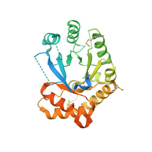
    Yersinia pestis remains a threat, with outbreaks of plague occurring in rural areas and its emergence as a weapon of bioterrorism; thus, an improved understanding of its various pathogenicity pathways is warranted. The rip (required for intracellular proliferation) virulence operon is required for Y. pestis survival in interferon-γ-treated macrophages and has been implicated in lowering macrophage-produced nitric oxide levels. RipC, one of three gene products from the rip operon, is annotated as a citrate lyase β subunit. Furthermore, the Y. pestis genome lacks genes that encode citrate lyase α and γ subunits, suggesting a unique functional role of RipC in the Y. pestis rip-mediated survival pathway. Here, the 2.45 Å resolution crystal structure of RipC revealed a homotrimer in which each monomer consists of a (β/α)(8) TIM-barrel fold. Furthermore, the trimeric state was confirmed in solution by size-exclusion chromatography. Through sequence and structure comparisons with homologous proteins, it is proposed that RipC is a putative CoA- or CoA-derivative binding protein.


  • Organizational Affiliation

    Department of Molecular Biology and Biochemistry, University of California, Irvine, CA 92697, USA.


Macromolecules
Find similar proteins by:  (by identity cutoff)  |  3D Structure
Entity ID: 1
MoleculeChains Sequence LengthOrganismDetailsImage
Citrate lyase
A, B, C
316Yersinia pestisMutation(s): 0 
Gene Names: citE2y2383YPO1928YP_1670
UniProt
Find proteins for Q9ZC38 (Yersinia pestis)
Explore Q9ZC38 
Go to UniProtKB:  Q9ZC38
Entity Groups  
Sequence Clusters30% Identity50% Identity70% Identity90% Identity95% Identity100% Identity
UniProt GroupQ9ZC38
Sequence Annotations
Expand
  • Reference Sequence
Small Molecules
Modified Residues  1 Unique
IDChains TypeFormula2D DiagramParent
MSE
Query on MSE
A, B, C
L-PEPTIDE LINKINGC5 H11 N O2 SeMET
Experimental Data & Validation

Experimental Data

  • Method: X-RAY DIFFRACTION
  • Resolution: 2.45 Å
  • R-Value Free: 0.256 
  • R-Value Work: 0.230 
  • R-Value Observed: 0.232 
  • Space Group: P 31 2 1
Unit Cell:
Length ( Å )Angle ( ˚ )
a = 88.056α = 90
b = 88.056β = 90
c = 197.021γ = 120
Software Package:
Software NamePurpose
DENZOdata reduction
SCALEPACKdata scaling
PHENIXrefinement
PDB_EXTRACTdata extraction
HKL-2000data collection
SOLVEphasing

Structure Validation

View Full Validation Report


Entry History 

Deposition Data

Revision History  (Full details and data files)

  • Version 1.0: 2012-01-11
    Type: Initial release
  • Version 1.1: 2012-03-28
    Changes: Database references
  • Version 1.2: 2017-11-08
    Changes: Refinement description
  • Version 1.3: 2018-05-30
    Changes: Data collection, Derived calculations