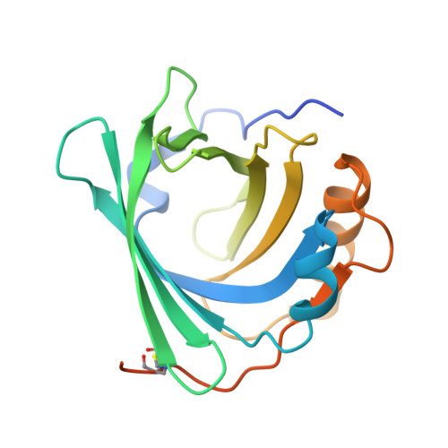
The crystal structure of human alpha(1)-microglobulin reveals a potential haem-binding site.
Meining, W., Skerra, A.(2012) Biochem J 445: 175-182
- PubMed: 22512701
- DOI: https://doi.org/10.1042/BJ20120448
- Primary Citation of Related Structures:
3QKG - PubMed Abstract:
We describe the 2.3 Å (1 Å=0.1 nm) X-ray structure of α1m (α1-microglobulin), an abundant protein in human blood plasma, which reveals the β-barrel fold typical for lipocalins with a deep pocket lined by four loops at its open rim. Loop #1 harbours the residue Cys34 which is responsible for covalent cross-linking with plasma IgA. A single disulfide bond between Cys72 and Cys169 connects the C-terminal segment to the β-barrel, as in many other lipocalins. The exposed imidazole side chains of His122 and His123 in loop #4 give rise to a double Ni2+-binding site together with a crystallographic neighbour. The closest structural relatives of α1m are the complement protein component C8γ, the L-prostaglandin D synthase and lipocalin 15, three other structurally characterized members of the lipocalin family in humans that have only distant sequence similarity. In contrast with these, α1m is initially expressed as a bifunctional fusion protein with the protease inhibitor bikunin. Neither the electron density nor ESI-MS (electrospray ionization MS) provide evidence for a chromophore bound to the recombinant α1m, also known as 'yellow/brown lipocalin'. However, the three side chains of Lys92, Lys118 and Lys130 that were reported to be involved in covalent chromophore binding appear to be freely accessible to ligands accommodated in the hydrophobic pocket. A structural feature similar to the well-known Cys-Pro haem-binding motif indicates the presence of a haem-binding site within the loop region of α1m, which explains previous biochemical findings and supports a physiological role in haem scavenging, as well as redox-mediated detoxification.
Organizational Affiliation:
Munich Center for Integrated Protein Science (CIPS-M) and Lehrstuhl für Biologische Chemie, Technische Universität München, Freising-Weihenstephan 85350, Germany.