3QHS

Crystal structure of full-length Hfq from Escherichia coli


Experimental Data Snapshot

  • Method: X-RAY DIFFRACTION
  • Resolution: 2.85 Å
  • R-Value Free: 0.250 
  • R-Value Work: 0.207 
  • R-Value Observed: 0.209 

wwPDB Validation   3D Report Full Report


This is version 1.2 of the entry. See complete history


Literature

Structural analysis of full-length Hfq from Escherichia coli

Beich-Frandsen, M.Vecerek, B.Sjoblom, B.Blasi, U.Djinovic-Carugo, K.

(2011) Acta Crystallogr Sect F Struct Biol Cryst Commun 67: 536-540

  • DOI: https://doi.org/10.1107/S174430911100786X
  • Primary Citation of Related Structures:  
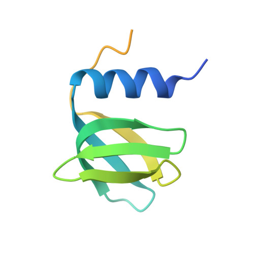
    3QHS

  • PubMed Abstract: 

    The structure of full-length host factor Qβ (Hfq) from Escherichia coli obtained from a crystal belonging to space group P1, with unit-cell parameters a = 61.91, b = 62.15, c = 81.26 Å, α = 78.6, β = 86.2, γ = 59.9°, was solved by molecular replacement to a resolution of 2.85 Å and refined to R(work) and R(free) values of 20.7% and 25.0%, respectively. Hfq from E. coli has previously been crystallized and the structure has been solved for the N-terminal 72 amino acids, which cover ~65% of the full-length sequence. Here, the purification, crystallization and structural data of the full 102-amino-acid protein are presented. These data revealed that the presence of the C-terminus changes the crystal packing of E. coli Hfq. The crystal structure is discussed in the context of the recently published solution structure of Hfq from E. coli.


  • Organizational Affiliation

    Department of Structural and Computational Biology, Max F. Perutz Laboratories, University of Vienna, Campus Vienna Biocenter 5, A-1030 Vienna, Austria.


Macromolecules
Find similar proteins by:  (by identity cutoff)  |  3D Structure
Entity ID: 1
MoleculeChains Sequence LengthOrganismDetailsImage
Protein hfq
A, B, C, D, E
A, B, C, D, E, F, G, H, I, J, K, L
102Escherichia coli K-12Mutation(s): 0 
Gene Names: b4172hfqJW4130
UniProt
Find proteins for P0A6X3 (Escherichia coli (strain K12))
Explore P0A6X3 
Go to UniProtKB:  P0A6X3
Entity Groups  
Sequence Clusters30% Identity50% Identity70% Identity90% Identity95% Identity100% Identity
UniProt GroupP0A6X3
Sequence Annotations
Expand
  • Reference Sequence
Experimental Data & Validation

Experimental Data

  • Method: X-RAY DIFFRACTION
  • Resolution: 2.85 Å
  • R-Value Free: 0.250 
  • R-Value Work: 0.207 
  • R-Value Observed: 0.209 
  • Space Group: P 1
Unit Cell:
Length ( Å )Angle ( ˚ )
a = 61.91α = 78.56
b = 62.15β = 86.16
c = 81.26γ = 59.86
Software Package:
Software NamePurpose
DNAdata collection
MOLREPphasing
REFMACrefinement
XDSdata reduction
XDSdata scaling

Structure Validation

View Full Validation Report


Entry History 

Deposition Data

Revision History  (Full details and data files)

  • Version 1.0: 2011-05-18
    Type: Initial release
  • Version 1.1: 2011-07-13
    Changes: Version format compliance
  • Version 1.2: 2023-11-01
    Changes: Data collection, Database references, Refinement description