3QGZ

Re-investigated high resolution crystal structure of histidine triad nucleotide-binding protein 1 (HINT1) from rabbit complexed with adenosine


Experimental Data Snapshot

  • Method: X-RAY DIFFRACTION
  • Resolution: 1.10 Å
  • R-Value Free: 0.168 
  • R-Value Work: 0.142 
  • R-Value Observed: 0.144 

wwPDB Validation   3D Report Full Report


Ligand Structure Quality Assessment 


This is version 1.4 of the entry. See complete history


Literature

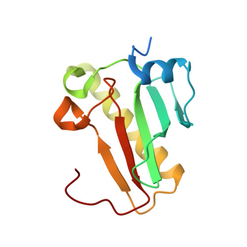
High-resolution X-ray crystal structure of rabbit histidine triad nucleotide-binding protein 1 (rHINT1) - adenosine complex at 1.10A resolution

Dolot, R.Ozga, M.Krakowiak, A.Nawrot, B.

(2011) Acta Crystallogr D Biol Crystallogr 67: 601-607

  • DOI: https://doi.org/10.1107/S0907444911015605
  • Primary Citation of Related Structures:  
    3QGZ

  • PubMed Abstract: 

    Histidine triad nucleotide-binding protein 1 (HINT1) represents the most ancient and widespread branch in the histidine-triad protein superfamily. HINT1 plays an important role in various biological processes and has been found in many species. Here, the first complete structure of the rabbit HINT1-adenosine complex is reported at 1.10 Å resolution, which is one of the highest resolutions obtained for a HINT1 structure. The final structure has an R(cryst) of 14.25% (R(free) = 16.77%) and the model exhibits good stereochemical qualities. A detailed analysis of the atomic resolution data allowed an update of the details of the protein structure in comparison to previously published data.


  • Organizational Affiliation

    Department of Bioorganic Chemistry, Centre of Molecular and Macromolecular Studies of the Polish Academy of Sciences, Łódź, Poland. rdolot@cbmm.lodz.pl


Macromolecules
Find similar proteins by:  (by identity cutoff)  |  3D Structure
Entity ID: 1
MoleculeChains Sequence LengthOrganismDetailsImage
Histidine triad nucleotide-binding protein 1126Oryctolagus cuniculusMutation(s): 0 
Gene Names: HINT1HINT
EC: 3
UniProt
Find proteins for P80912 (Oryctolagus cuniculus)
Explore P80912 
Go to UniProtKB:  P80912
Entity Groups  
Sequence Clusters30% Identity50% Identity70% Identity90% Identity95% Identity100% Identity
UniProt GroupP80912
Sequence Annotations
Expand
  • Reference Sequence
Small Molecules
Ligands 1 Unique
IDChains Name / Formula / InChI Key2D Diagram3D Interactions
ADN
Query on ADN

Download Ideal Coordinates CCD File 
B [auth A]ADENOSINE
C10 H13 N5 O4
OIRDTQYFTABQOQ-KQYNXXCUSA-N
Experimental Data & Validation

Experimental Data

  • Method: X-RAY DIFFRACTION
  • Resolution: 1.10 Å
  • R-Value Free: 0.168 
  • R-Value Work: 0.142 
  • R-Value Observed: 0.144 
  • Space Group: P 43 21 2
Unit Cell:
Length ( Å )Angle ( ˚ )
a = 39.747α = 90
b = 39.747β = 90
c = 141.83γ = 90
Software Package:
Software NamePurpose
MAR345data collection
REFMACrefinement
MOSFLMdata reduction
SCALAdata scaling
REFMACphasing

Structure Validation

View Full Validation Report


Ligand Structure Quality Assessment 


Entry History 

Deposition Data

Revision History  (Full details and data files)

  • Version 1.0: 2011-02-16
    Type: Initial release
  • Version 1.1: 2011-07-13
    Changes: Version format compliance
  • Version 1.2: 2011-10-26
    Changes: Database references
  • Version 1.3: 2017-11-08
    Changes: Data collection, Refinement description
  • Version 1.4: 2023-09-13
    Changes: Data collection, Database references, Derived calculations, Refinement description