3QE3

Sheep liver sorbitol dehydrogenase


Experimental Data Snapshot

  • Method: X-RAY DIFFRACTION
  • Resolution: 1.90 Å
  • R-Value Free: 0.234 
  • R-Value Work: 0.192 
  • R-Value Observed: 0.195 

wwPDB Validation   3D Report Full Report


This is version 1.3 of the entry. See complete history


Literature

X-ray crystal structure and small-angle X-ray scattering of sheep liver sorbitol dehydrogenase.

Yennawar, H.Moller, M.Gillilan, R.Yennawar, N.

(2011) Acta Crystallogr D Biol Crystallogr 67: 440-446

  • DOI: https://doi.org/10.1107/S0907444911007815
  • Primary Citation of Related Structures:  
    3QE3

  • PubMed Abstract: 

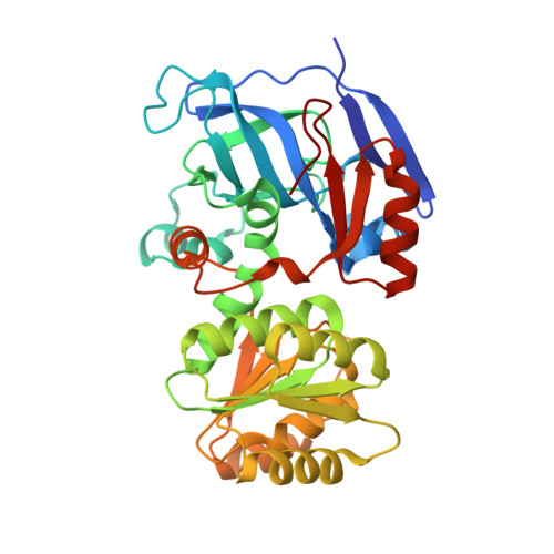
    The X-ray crystal structure of sheep liver sorbitol dehydrogenase (slSDH) has been determined using the crystal structure of human sorbitol dehydrogenase (hSDH) as a molecular-replacement model. slSDH crystallized in space group I222 with one monomer in the asymmetric unit. A conserved tetramer that superposes well with that seen in hSDH (despite belonging to a different space group) and obeying the 222 crystal symmetry is seen in slSDH. An acetate molecule is bound in the active site, coordinating to the active-site zinc through a water molecule. Glycerol, a substrate of slSDH, also occupies the substrate-binding pocket together with the acetate designed by nature to fit large polyol substrates. The substrate-binding pocket is seen to be in close proximity to the tetramer interface, which explains the need for the structural integrity of the tetramer for enzyme activity. Small-angle X-ray scattering was also used to identify the quaternary structure of the tetramer of slSDH in solution.


  • Organizational Affiliation

    Department of Biochemistry and Molecular Biology and Huck Institutes of Life Sciences, Pennsylvania State University, 8 Althouse Laboratory, University Park, PA 16802, USA.


Macromolecules
Find similar proteins by:  (by identity cutoff)  |  3D Structure
Entity ID: 1
MoleculeChains Sequence LengthOrganismDetailsImage
Sorbitol dehydrogenase355Ovis ariesMutation(s): 0 
Gene Names: SORD
EC: 1.1.1.14
UniProt
Find proteins for P07846 (Ovis aries)
Explore P07846 
Go to UniProtKB:  P07846
Entity Groups  
Sequence Clusters30% Identity50% Identity70% Identity90% Identity95% Identity100% Identity
UniProt GroupP07846
Sequence Annotations
Expand
  • Reference Sequence
Experimental Data & Validation

Experimental Data

  • Method: X-RAY DIFFRACTION
  • Resolution: 1.90 Å
  • R-Value Free: 0.234 
  • R-Value Work: 0.192 
  • R-Value Observed: 0.195 
  • Space Group: I 2 2 2
Unit Cell:
Length ( Å )Angle ( ˚ )
a = 67.749α = 90
b = 85.881β = 90
c = 119.888γ = 90
Software Package:
Software NamePurpose
CrystalCleardata collection
PHENIXmodel building
PHENIXrefinement
HKL-2000data reduction
HKL-2000data scaling
PHENIXphasing

Structure Validation

View Full Validation Report


Entry History 

Deposition Data

Revision History  (Full details and data files)

  • Version 1.0: 2011-04-27
    Type: Initial release
  • Version 1.1: 2011-07-13
    Changes: Version format compliance
  • Version 1.2: 2013-03-27
    Changes: Database references
  • Version 1.3: 2023-09-13
    Changes: Data collection, Database references, Derived calculations, Refinement description